Amen Clinics Position on Chronic Traumatic Encephalopathy (CTE)
By Daniel G. Amen, MD
Chronic traumatic encephalopathy (CTE) was brought into the public’s awareness largely through the movie Concussion, starring Will Smith, based on the work of neuropathologist Bennet Omalu. When he did the autopsy on Pittsburgh Steeler Hall of Fame Center Mike Webster, he noticed something very different in his brain that had not been reported. There were excessive deposits of an abnormal protein in his brain, called tau. He subsequently noticed this pattern in other deceased NFL players who struggled with severe cognitive and emotional issues.
‘I am interested in the living and doing everything I can to delay their journey to the autopsy table.’ — Daniel G. Amen, MD

 In brain SPECT images, a healthy scan shows full, even, symmetrical blood flow. The “holes” in AD’s pre-treatment scan represent areas of low blood flow. His follow-up brain scan shows healthier overall blood flow.
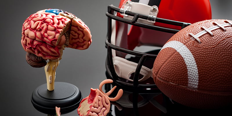
INSIDE THE CTE BRAIN
Tau protein is essential as it provides the lattice-like structure of brain cells. However, when the brain is damaged from repeated concussions, common in football and soccer players, tau breaks down and pierces through cell membranes, causing an inflammatory response that damages the brain. I was blessed to be a consultant on the Concussion. Since CTE can only be diagnosed with certainty on autopsied slices of brain tissue, it can only be diagnosed after death. I am friends with Dr. Omalu, and we have published scientific papers together. I tell him, his patients are the dead, but I am interested in the living and doing everything I can to delay their journey to his autopsy table.WHY HEAD TRAUMA ISN’T HOPELESS
Yet, the lore in medical circles is that CTE is permanent, progressive, and untreatable. I think that is nonsense and there is virtually no scientific evidence to support the lack of hope. Yet, because many people believe it, former football players and other professional athletes avoid getting help because they do not believe there’s anything they can do to stem the inevitable destruction of their brains. Some in the media have accused me of giving people with CTE false hope. Let me be clear, the scans we do at Amen Clinics do not diagnose CTE (you need a pathology slide for that). However, there is large body of peer-reviewed scientific research demonstrating that brain SPECT imaging can diagnose the current impact of past concussions, and whether or not with proper help, the brain can improve. The media tends to be purveyors of doom and despair, because that sells papers or gets more eyeballs for advertisers, but my 40-year career has shown that there is reason to be optimistic about the brain’s ability to heal. Here’s an example. In July 2007, College Football Hall of Fame running back from USC and former professional football player Anthony Davis (AD), came to see me at Amen Clinics because of problems with memory, periods of confusion, and irritability. A professor at USC told him about our work, and he thought we could help him. He was also concerned about the cognitive problems he saw in other retired football players and was hoping to find a way to help them too. At 54, Anthony’s brain looked like he was 85. It showed clear evidence of brain trauma to the prefrontal cortex and left temporal lobe. I put AD on a brain rehabilitation program described below, and within several months he told me that he felt better, was more focused, and had better energy and a sharper memory. Through my relationship with AD, in 2009 I partnered with the Los Angeles Chapter of the Retired NFL Players Association, which co-sponsored the largest brain imaging and rehabilitation study on active and retired NFL players. At the time, the NFL was still saying they did not know if playing football caused long-term brain damage, but they had never done any brain-imaging studies on players to prove it or not. My colleagues and I decided to tackle the issue. To date, we have scanned and treated more than 300 active and retired players using SPECT, a functional brain-imaging technique that measures blood flow and activity in the brain. The pre-treatment brain scans showed high levels of damage in almost all of the players. Damage was seen most commonly in the following regions:- Prefrontal cortex—involved with judgment, planning, forethought, and impulse control
- Temporal lobes—involved with learning, memory, and mood stability
- Cerebellum—involved with mental agility and processing speed
AD’s Brain Before and After 10 Years
In brain SPECT images, a healthy scan shows full, even, symmetrical blood flow. The “holes” in AD’s pre-treatment scan represent areas of low blood flow. His follow-up brain scan shows healthier overall blood flow.