Brain SPECT Made Ridiculously Simple: Part 3
Brain SPECT imaging has dramatically influenced the way we diagnose and treat our patients. In Part 1 of this blog, you learned the basics about brain SPECT imaging and why it is considered the future of psychiatry. In Part 2, you discovered some of the common patterns seen in SPECT brain scans. In this part, you will discover three additional brain SPECT imaging patterns, what causes them, and the corresponding interventions.
Brain SPECT Imaging Pattern 4: Hyperfrontality (frontal lobes work too hard)
SPECT Scan: Hyperfrontality Examples
OCD: Hyperfrontal pattern
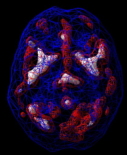
Anxiety with obsessive thoughts: Hyperfrontal pattern
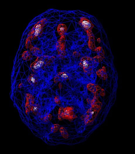
In this pattern, the frontal lobes of the brain are working too hard. We saw this pattern in a 37-year-old woman who came to see us because she was having trouble in her relationship and at work. Her husband complained that she was very negative and argumentative. At work, whenever something didn’t go as planned, she would stew about it for hours and miss deadlines. We diagnosed her with overfocused anxiety and depression and treated her with a combination of exercise, supplements, and specific foods. The treatment helped her relax, feel happier, and be more flexible in all areas of her life. Hyperfrontality can be seen in the following conditions:
• Obsessive-Compulsive disorder spectrum
• Oppositional defiant disorder
• Autism spectrum
• People who get stuck, worried, rigid, inflexible (when things get upset is things don’t go their way)
• Overfocused depression or anxiety (can’t stop thinking bad thoughts)
Interventions:
Increase serotonin to calm the brain through the following:
• Exercise
• Diet
• Supplements (5HTP or saffron)
• SSRIs (antidepressant medication, if necessary)
Brain SPECT Imaging Pattern 5: Hypofrontality (low frontal lobe activity)
SPECT Scan: Hypofrontality
This pattern shows decreased activity in the front part of the brain

Hypofrontality indicates low activity in the frontal lobes. We noticed this pattern in a 7-year-old boy, who was restless, fidgety, unable to focus, and doing poorly in school. While he was at rest, his brain scan showed full even activity, but when he performed a concentration task his frontal lobes decreased in activity—an indicator of ADHD. In people who don’t have ADHD, the frontal lobes typically increase in activity while concentrating. With the right diagnosis and interventions, the boy was better able to pay attention in class and his grades improved. Hypofrontality is associated with:
• ADHD
• Schizophrenia
• Traumatic brain injury
• Medications
• Predicts relapse in alcoholics
• Lack of conscientiousness
• Lack of forward thinking
• Some forms of depression
Interventions:
• Exercise
• Stimulating supplements, such as green tea, l-tyrosine, and rhodiola
• Stimulants, if ADHD
• Stimulating antipsychotics, if needed
• Stimulating antidepressants or SAMe, if depressed
Brain SPECT Imaging Pattern 6: Temporal Lobe Abnormalities
SPECT Scans: Temporal Lobe Examples
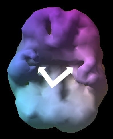
Intermittent Explosive Disorder: Low prefrontal and left and right temporal lobe activity (arrows)
Social Skills Issues, trouble reading social cues: Low right temporal lobe activity
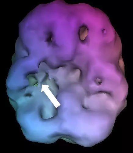
This pattern indicates one or both of the temporal lobes are unbalanced. We saw this pattern in a 23-year-old man, who came to see us when his employer told him he would be fired if he didn’t get his anger under control. The young man would suddenly lash out at coworkers for no reason and then go back to being pleasant. After seeing temporal abnormalities in his SPECT brain scan, he began hyperbaric oxygen therapy and started following a ketogenic diet, which helped him gain control of his anger. Not only did he keep his job, he got promoted to a managerial role and gained the trust of his colleagues. Temporal lobe abnormalities can be seen in:
• Temporal lobe epilepsy
• Temporal lobe dysrhythmia
• Dyslexia
• Mood instability
• Irritability
• Intermittent explosive disorder
Interventions:
• Ketogenic diet
• Hyperbaric oxygen therapy (HBOT)
• Anti-seizure medications
• Memory-enhancing medications or supplements, if memory is the problem
How does identifying these brain SPECT imaging patterns influence treatment?
In a 2012 study we conducted, we asked 7 psychiatrists to evaluate 109 consecutive charts without scans and then with scans. In 8 times out of 10, adding the scan into the review changed the diagnosis and/or treatment. In over 1 in 5 cases, the scan revealed an unexpected brain injury, and in another 1 in 5 cases, it revealed unexpected toxicity. And 60 percent of the time, it changed the medications or supplements recommended.
With brain SPECT imaging, do patients get better faster?
This is one of the most common questions I hear. We do outcome studies on all of our patients—we have over 7,000 outcomes on our patients. At the end of 6 months, if they are treated at Amen Clinics 84 percent of our complicated patients report being improved. And quality of life scores go up in 85 percent of patients. You can change your brain and change your life.
The Amen Clinics has been using brain SPECT imaging with its patients since 1991 and has the world’s largest functional brain imaging database related to brain health/mental health issues with over 150,000 SPECT scans on patients from 120 countries.
If you or someone you know would like more information about brain SPECT imaging at Amen Clinics, watch these two videos, or to make an appointment, visit us online or call: 888-288-9834.
SPECT Made Ridiculously Simple with Dr. Daniel Amen Part 1
SPECT Made Ridiculously Simple with Dr. Daniel Amen Part 2