
TL;DR
Autism has a strong genetic component, but there is no single “autism gene” or one clear cause. Research suggests that inherited genetic factors, along with environmental and developmental influences during early brain development, may all play a role.
Autism is also highly individual. People on the spectrum can have different strengths, challenges, communication styles, and support needs. Co-occurring conditions such as anxiety, ADHD, sleep problems, or gastrointestinal issues may also influence how symptoms appear.
At Amen Clinics, brain SPECT imaging is used as part of a comprehensive evaluation to help clinicians better understand brain function and contributing factors. This added information supports precision medicine, allowing for more accurate diagnoses and highly personalized treatment plans.
Treatment recommendations are guided by holistic psychiatry, which focuses on caring for the whole person through lifestyle strategies, nutrition, supplements, therapy, and medication when necessary.
Medically reviewed by Kandace Licciardi, MD, Medical Director, Amen Clinics

If you’re a parent, caregiver, or someone trying to better understand autism, you’ve probably asked the question: Is autism genetic?
The short answer is yes, genetics appear to play a major role in autism. But the full answer is far more complex, and more hopeful, than many people realize.
Research shows that autism spectrum disorder (ASD) is strongly linked to inherited genetic factors. At the same time, scientists also believe that early brain development, environmental influences, and biological factors may affect how autism traits appear and develop over time. In other words, autism is not caused by one single gene or one single event.
Understanding this matters because autism is not a character flaw, a parenting failure, or a one-size-fits-all condition. Every person on the spectrum has a unique brain, unique strengths, and unique challenges.
In this blog, you’ll learn what current science says about the genetic roots of autism, whether autism is hereditary, how environmental factors may influence risk, and how Amen Clinics approaches autism through a personalized, brain-health-focused perspective.
Related: The Many Faces of Autism
Is autism genetic? No single genetic case is clearly identified with autism. The risk of autism more likely involves multiple genes that are working together alongside complex biological pathways that may affect brain development.
Autism spectrum disorder is a neurodevelopmental condition that can influence brain function, shaping how an individual communicates, interacts socially, and understands or even responds to information.
Autism is fairly common. According to the Centers for Disease Control and Prevention (CDC), about 1 in every 31children in the U.S. is diagnosed with ASD.
According to the World Health Organization, about 1 in 127 people globally are on the autism spectrum, which underscores how widely it affects populations around the world.
Research shows that autism has a strong genetic component, but it is not usually considered a single-gene disorder.
Some people with autism also have known genetic conditions or identifiable genetic differences. According to the CDC certain individuals with ASD have genetic conditions linked to autism, while many others do not have one clear genetic explanation.
Part of the confusion comes from the term “genetic disorder,” which can mean different things. Some genetic disorders are caused by a mutation in a single gene. Others involve many genes and complex biological pathways working together.
Autism is generally understood as a neurodevelopmental condition influenced by multiple genes, rather than one specific “autism gene.” In fact, researchers believe that many different genetic variations may contribute to autism risk in different people.
That’s why autism is described as highly genetic, but also highly complex. Genetics may strongly influence autism, yet there is no single genetic cause that explains every person on the spectrum.
Sometimes, autism may be connected to particular genetic syndromes. For instance, according to research, conditions like Down syndrome or Fragile X syndrome have been known to increase the chances of autism.
For many, however, no single genetic case is clearly identified with ASD. The risk of autism more likely involves multiple genes that are working together alongside complex biological pathways that may affect brain development.
That explains why autism is usually described as polygenetic, which means it can be influenced by the combined influence of multiple genes and not a single or specific gene.
Autism is not believed to be caused by genetics alone or environment alone.
The CDC suggests that both likely play a role in early brain development and that there is no single cause of autism. Instead, a combination of genetic, biological, and environmental factors may contribute to autism risk.
The National Institute of Mental Health (NIMH) also supports the understanding that with ASD, inherited genes along with certain environmental influences may affect how the brain develops during pregnancy and early childhood.
This does not mean that one specific environmental factor causes autism. Rather, experts believe autism develops through a complex interaction of many influences over time.
Some people inherit genetic differences that can increase the chances of developmental traits that are related to autism. Those gene factors may affect the formation and functioning of the brain, which creates a certain level of vulnerability; however, they don’t determine outcomes on their own.
Researchers believe that environmental influences during pregnancy and early childhood may interact with inherited genetic risk factors. This is a critical stage of development because the brain is growing and changing rapidly.
According to NIMH and the National Institute of Child Health and Human Development (NICHD), autism likely develops through a combination of genetic, biological, and environmental influences that work together over time to shape early brain development.
Risk factors are different from direct causes. An environmental association doesn’t imply that the autism in a particular child was caused by a specific factor. Researchers believe autism risk is influenced by a combination of factors that affect early brain development.
Researchers continue to study how non-genetic influences may affect autism risk, especially in people who are already genetically susceptible. The NIMH lists factors associated with increased likelihood, including having older parents and having a very low birth weight. The CDC also reports that environmental and biologic factors may make a child more likely to have ASD.
These factors can’t directly cause autism but they are part of a broader pattern of influences that researchers are still working to understand. Environmental factors may have a role in the development of the risk of autism, but it seems like they work within a larger picture, including early brain development and genetics.
Oversimplified explanations about autism and genetics can create confusion, stress, and unnecessary guilt for families. Research does not support the idea that autism is caused only by genes or only by environmental factors.
It is also important not to assume that every autistic person has the same underlying biology or developmental pathway. Experts continue to study the many factors that may influence brain development, and understanding is still evolving.
Another harmful misconception is blaming parents or caregivers for autism. Autism is not caused by parenting choices, and framing it that way can leave families feeling blamed instead of supported.
Families deserve clear, balanced information rooted in science and compassion. Autism is a neurodevelopmental condition, not a character flaw or a reflection of bad parenting.
Autism is called a spectrum condition because it can look very different from one person to another. Individuals on the spectrum may have different strengths, challenges, communication styles, and support needs.
According to NIMH, autism can affect social interaction, communication, behavior, and learning. However, the way these traits appear, and how strongly they affect daily life, varies widely.
Some people with autism may need significant day-to-day support, while others live highly independent lives but still experience challenges related to sensory processing, social communication, attention, or mood.
This variation is important when it comes to evaluation and treatment. Having treating more than 100,000 patients, clinicians at Amen Clinics have observed that many children and adults with autism also experience co-occurring medical or mental health conditions, such as anxiety, ADHD, sleep problems, or mood issues, which can influence how symptoms appear.
In some cases, brain SPECT imaging may be used as part of a broader evaluation to help clinicians better understand individual patterns of brain activity related to behavior, attention, learning, and emotional regulation.
These differences are one reason personalized assessment and individualized support strategies are so important for people on the autism spectrum.
Children and adults with autism may also experience other medical or mental health conditions that affect daily functioning and influence how symptoms appear.
From more than 35 years of clinical experience at Amen Clinics, we have seen that co-occurring conditions such as anxiety, ADHD, epilepsy, sleep problems, and gastrointestinal issues are common in people on the spectrum. These issues can affect mood, attention, behavior, learning, and overall quality of life.
That’s why it is important not to assume every challenge is caused by autism alone. Identifying co-occurring conditions can provide a clearer understanding of what a person is experiencing and help guide more effective support and treatment strategies.
Personalized care is especially important because each individual may have a different combination of strengths, challenges, and underlying health concerns.
Traditional approaches to autism often focus primarily on diagnosing symptoms and managing behaviors. While these strategies can be helpful, they do not always address the underlying differences in how each person’s brain functions.
At Amen Clinics, care is guided by a brain-health-focused approach that looks beyond symptoms to better understand what may be affecting mood, behavior, learning, attention, and daily functioning.
With a comprehensive evaluation at Amen Clinics, our clinicians use brain SPECT imaging to examine patterns of blood flow and brain activity, which helps them identify co-occurring issues that may be influencing symptoms. This approach recognizes that two people with the same diagnosis may have very different brain function patterns and support needs.
We practice holistic psychiatry, which means we combine targeted nutrition, supplements, therapy, lifestyle strategies, and medication when appropriate. It is a whole-body approach that supports the mind and body.
By identifying individual brain patterns and related health concerns, clinicians can develop more personalized treatment plans designed to support each person’s unique needs and strengths.
Single photon emission computed tomography (SPECT) imaging looks at blood flow and activity patterns in the brain, offering insight into how the brain functions. This differs from scans such as CT or MRI, which primarily show brain structure. By focusing on function, brain SPECT imaging can help identify areas of the brain that may be underactive or overactive.
At Amen Clinics, brain SPECT imaging is used as part of a comprehensive evaluation that may also include a detailed personal history, symptom assessment, neuropsychological testing, and lab work when appropriate. This broader approach helps clinicians better understand factors that may be affecting mood, attention, learning, behavior, and overall functioning.
Drawing from decades of experience and one of the world’s largest databases of brain scans, Amen Clinics uses these insights to support precision medicine and more individualized care plans for people on the autism spectrum.
Our whole-body approach to holistic psychiatry combines brain-based insights with targeted nutrition, supplements, therapy, lifestyle strategies, and medication when needed. Rather than relying on a one-size-fits-all approach, treatment is tailored to the unique needs of each individual.
If developmental, behavioral, social, or communication challenges are beginning to interfere with daily life, it may be helpful to seek a professional evaluation.
It is especially important to look for support when concerns go beyond what can be managed through school accommodations or self-help strategies alone, or when there are additional issues such as anxiety, sleep problems, attention difficulties, or other medical concerns.
An evaluation can also help if you are unsure whether autism fully explains what you or your child is experiencing, or if you want a clearer understanding of strengths, challenges, and support needs.
Early and accurate autism diagnosis is critical. The CDC reports that early intervention can improve outcomes for children with autism, which is why it is important to seek guidance when concerns arise. In a study published in Autism Research, it was found that 25 percent of children with autism under the age of 8 fail to get diagnosed, especially in minority populations.
A comprehensive evaluation can help identify whether autism, co-occurring conditions, or other factors may be contributing to symptoms and daily functioning.
Autism is strongly genetic, but it is not only genetic. The best current evidence points to a mix of inherited risk and environmental or developmental influences that affect early brain development.
The goal is not just to ask whether autism is genetic. It is to better understand the individual person, identify co-occurring challenges, and build support around what their brain and body need most.
If you are trying to understand autism more clearly and want a more personalized view of what may be affecting brain function, a comprehensive evaluation may help you find more useful answers.
Genetics play a major role in autism, which means autism can run in families. However, there is not one single “autism gene,” and inherited risk can involve many different genes working together.
Current research does not support the idea that one environmental factor directly causes autism. Researchers believe environmental influences may interact with genetic factors during early brain development.
Autism is a spectrum condition, so strengths, challenges, communication styles, and support needs can vary widely. Some people may need significant daily support, while others live independently but still experience social, sensory, or behavioral challenges.
At Amen Clinics, brain SPECT imaging is used as part of a comprehensive evaluation that may also include medical history, symptom assessment, cognitive testing, and other clinical tools. This allows our clinicians to practice precision medicine, rendering more accurate diagnoses, especially when there may be co-occurring conditions, and more targeted, personalized treatment plans.
Plans are guided by holistic psychiatry, which takes a whole-body approach. They prioritize natural ways to treat mental health conditions and and incorporate lifestyle recommendations (diet, sleep, supplements, exercise, etc.), therapy, and medication when necessary.
|
Autism spectrum disorder and other mental health conditions can’t wait. At Amen Clinics, we practice precision medicine—using brain SPECT imaging and comprehensive evaluations to understand what’s really happening in your brain, not just your symptoms. Our whole-body approach to holistic psychiatry combines cutting-edge neuroscience with natural ways to treat mental health conditions, including targeted nutrition, supplements, lifestyle strategies, therapy, and medications (when necessary). Every treatment plan is personalized to address the root causes of your struggles and support the health of your brain, body, and mind. Don’t settle for guesswork. You deserve answers—and a plan built specifically for you. Speak with a Brain Health Advisor today at 888-288-9834 or visit our contact page to get started. |
About the Reviewer
Kandace Licciardi, MD, Medical Director, Amen Clinics
Dr. Kandace Licciardi is a double board-certified psychiatrist in adult and forensic psychiatry and serves as Medical Director of Amen Clinics. She takes a comprehensive, whole-person approach to care, integrating evidence-based psychiatric treatment with lifestyle, social, and wellness factors. Dr. Licciardi has extensive experience treating depression, anxiety, bipolar disorder, psychotic disorders, and borderline personality disorder, with special expertise in early psychosis intervention. She completed her psychiatry residency at Maimonides Medical Center and her forensic psychiatry fellowship at New York University.
La Monica, I., Di Iorio, M. R., Sica, A., Rufino, F., Sotira, C., Pastore, L., & Lombardo, B. (2025). Autism spectrum disorder: genetic mechanisms and inheritance patterns. Genes, 16(5), 478. doi: 10.3390/genes16050478
El-Fishawy, P., & State, M. W. (2010). The genetics of autism: key issues, recent findings and clinical implications. The Psychiatric Clinics of North America, 33(1), 83. https://doi.org/10.1016/j.psc.2009.12.002
Lorang E, Sterling A; Expressive Language Consortium. The impact of autism spectrum disorder symptoms on gesture use in fragile X syndrome and Down syndrome. Autism Dev Lang Impair. 2017 Jan-Dec;2:10. https://pmc.ncbi.nlm.nih.gov/articles/PMC6191055/
Centers for Disease Control and Prevention. (n.d.). Autism spectrum disorder (ASD): About autism spectrum disorder. https://www.cdc.gov/autism/about/index.html
National Institute of Mental Health. (n.d.). Autism spectrum disorder. U.S. Department of Health and Human Services, National Institutes of Health. https://www.nimh.nih.gov/health/topics/autism-spectrum-disorders-asd
Centers for Disease Control and Prevention. (n.d.). Autism spectrum disorder: Accessing services. U.S. Department of Health and Human Services. https://www.cdc.gov/autism/treatment/accessing-services.html
Wiggins LD, Durkin M, Esler A, Lee LC, Zahorodny W, Rice C, Yeargin-Allsopp M, Dowling NF, Hall-Lande J, Morrier MJ, Christensen D, Shenouda J, Baio J. Disparities in Documented Diagnoses of Autism Spectrum Disorder Based on Demographic, Individual, and Service Factors. Autism Res. 2020 Mar;13(3):464-473. doi: 10.1002/aur.2255. Epub 2019 Dec 23. PMID: 31868321; PMCID: PMC7521364.
Centers for Disease Control and Prevention. (n.d.). Data and research on autism spectrum disorder. U.S. Department of Health and Human Services. https://www.cdc.gov/autism/data-research/index.htm
World Health Organization. (2025, September 17). Autism spectrum disorders. https://www.who.int/westernpacific/newsroom/fact-sheets/detail/autism-spectrum-disorders
Sleep is not a break from brain activity. It’s when your brain does some of its most important work.
While you sleep, your brain cleans itself, repairs damaged cells, consolidates memories, and resets the systems that control mood, focus, judgment, and emotional balance. This nightly renewal process helps wash away toxic metabolic waste, such as amyloid plaques linked to Alzheimer’s disease, and supports long-term brain health and longevity.
Consistently getting quality sleep is essential for mental health. Without enough restorative sleep, the brain can’t fully recharge. Memory suffers. Emotions become harder to regulate. Stress tolerance drops. And over time, disrupted sleep can quietly erode the very brain circuits responsible for clear thinking, impulse control, and emotional stability.
Chronic sleep deprivation doesn’t just make you tired. It increases irritability, worsens anxiety and depression, impairs concentration, and makes everyday stress feel overwhelming. When poor sleep continues long enough, the brain’s ability to function optimally begins to break down.
In this blog, we’ll explore how sleep affects mental health, what happens in the brain when sleep is disrupted, and why protecting your sleep may be one of the most powerful ways to protect your brain.
Medically reviewed by Kandace Licciardi, MD, Medical Director, Amen Clinics
Related: How Do Sleep Disorders Impact the Brain and Mental Health?

If you really want to know how sleep affects mental health, you must first understand what happens inside your brain while you sleep.
During sleep, your brain processes emotional experiences, resets your stress response systems, and strengthens memory and learning. This nightly reset supports emotional balance, clearer thinking, and improved focus.
Studies show that when you have stable sleep patterns, they help support more balanced moods. Stable sleeping patterns also increase your resilience to stress.
In the long run, insufficient or disrupted sleep interferes with these key brain functions, which can make it harder for you to regulate your emotions and manage daily stressors.
While you sleep, your brain cleans itself, repairs damaged cells, consolidates memories, and resets the systems that control mood, focus, judgment, and emotional balance
Lack of sleep interferes with your mental health by disrupting the brain pathways that are responsible for stress resilience, cognitive control, and emotional regulation. Knowing this, you probably imagine how lack of sleep affects mental health in the long run. It’s not good.
When you fragment or reduce your sleep consistently, your mental performance and emotional balance begin to decline. Over time, such changes may affect how you think, feel, and respond to the challenges you experience every day. You may experience any of the following mental health effects:
Insufficient sleep can also affect the key brain regions that are involved in emotional regulation and impulse control, especially in the areas that support self-control and judgment. When those regions are affected, it may be harder for you to manage emotional reactions.
Looking at the brain’s response to ongoing sleep loss tells us a lot about sleep and mental health. When you are experiencing long-term sleep loss, your brain begins to have changes in the regions involved in judgment, emotional control, and focus.
One of the commonly affected regions is the prefrontal cortex. This area of the brain plays a key role in decision-making, planning, and attention. Research associates ongoing sleep deprivation with altered blood flow in the prefrontal cortex. Reduced blood flow is associated with poor brain function. This might explain why judgment, concentration, and impulse control may feel more difficult for individuals whose sleep is consistently limited.
Your amygdala (the region in your brain that is involved in detecting potential threats and processing emotions) can become overactive if you don’t get sufficient sleep. An overactive amygdala may cause you to experience stronger emotional responses, like heightened anxiety, increased irritability, or feeling overwhelmed emotionally in situations that would otherwise feel manageable.
Brain imaging has helped experts clearly see how chronic sleep problems affect the brain. At Amen Clinics, brain SPECT imaging shows disrupted blood-flow activity patterns in individuals with long-term sleep deprivation. SPECT scans consistently reveal that insufficient sleep alters brain function in ways that undermine mood regulation, focus, and emotional control.
Related: How to Improve Sleep Quality
When considering how lack of sleep impacts your mental health, it’s important to examine how your brain handles stress management, emotional regulation, and cognitive performance.
Consistently experiencing insufficient or disrupted sleep can influence the symptoms that are commonly linked to a number of mental health conditions. Inadequate sleep shapes how your brain processes mood, stress, and inflammation, which may impact your mental health. Here are some of the mental health symptoms and conditions that can result from too much disrupted or inadequate sleep.
Lack of adequate sleep can heighten your body’s stress response and make your brain more reactive to perceived threats. If the brain doesn’t get restorative rest, it can become more sensitive to everyday stressors and anxiety triggers. With such an increase in reactivity, it can be difficult for you to feel grounded, emotionally steady, or calm.
Over time, poor sleep has been known to predict or worsen depressive symptoms. Difficulties in sleeping usually appear before an individual experience changes in mood. The same difficulties can also intensify the feelings of reduced motivation, low energy, and emotional heaviness. This is a two-way relationship that helps to explain why, when you improve your sleep quality, you support your mood stability.
Insufficient sleep reduces working memory, attention control, and the ability to initiate different tasks. Lack of sufficient sleep causes the brain’s executive functions to become less efficient, which makes it harder for individuals to follow through with responsibilities or to stay focused. Such effects may amplify or resemble challenges in mental focus and organization.
With disrupted sleep, it can be difficult for your brain to regulate emotional responses or process memories. During deep restorative sleep, the brain works to reduce emotional intensity and integrate emotional experiences over time. This process can be less effective, especially for someone whose sleep is disrupted, which can eventually contribute to difficulties in managing distress and heightened emotional responses. This is especially true if an individual with PTSD or emotional trauma encounters a trigger.
Sleep problems usually escalate because of reinforcing one another in a repeating loop. When you’ve had insufficient sleep, your body and brain become more stressed. This heightened stress makes it even more difficult for you to sleep the following night.
The loss of sleep causes an increase in your stress levels by activating the stress response systems in your brain. When stress is elevated, it then interferes with your brain’s ability to rest and transition into relaxing sleep. This leads to further sleep disruption. After some time, this becomes a cycle in which heightened stress and poor sleep feed into each other continuously.
Anxiety and rumination tend to increase at night. This is when the external distractions quiet down and internal thoughts become louder.
If you’ve not been having adequate sleep, it means that your brain may experience difficulty trying to quiet the worry, replaying concerns, and anticipating future stressors. Such mental activity can cause a delay in sleep onset or fragmented sleep throughout the night.
While this pattern continues, your brain can become restless and overstimulated instead of calm and prepared for rest. Your neural activity remains elevated instead of winding down. This makes sleep feel shallow and difficult to sustain. Such an ongoing stimulation can further reinforce the sleep-stress cycle.
Emotional exhaustion can add another layer to these challenges. When your emotional and mental energy are depleted, it can be more difficult for you to maintain the healthy habits that enhance effective sleeping patterns, such as stress management, consistent routines or wind-down practices. That makes it more challenging to break the cycle even when you have a strong desire for better sleep.
When you are having sleep problems, the impact usually shows up in the way you feel, think, and respond to the situations you come across every day. At first, some of these challenges may be hard to discern. However, if you know what to be aware of, you might better be able to recognize them. They are clear indicators that your sleep has been affecting your mental health. Here are some of the signs to look out for:
To improve your sleep, consider making small, consistent changes that can support the way your brain regulates focus, stress, and mood. Science-backed sleep habits can help regulate your nervous system and support your overall mental well-being in the long run. Here are some simple habits to embrace:
Maintain Consistent Sleep and Wake times
When you choose to sleep and wake up at the same time every day, your internal clock begins to regulate. Doing it consistently helps support better focus, a more stable mood, and improved resilience during stressful experiences.
Limit Screen Time Before Bed
Screen time is known to keep the brain mentally alert when it’s supposed to be winding down, according to research. If you can conclude screen time at least an hour before going to bed, you’ll help your brain more easily transition into rest.
Create a Quiet, Dark Sleeping Environment
Excess noise and light can interfere with the quality of your sleep, no matter how long you stay asleep. A dark, calm space will help to ensure that your brain remains in deeper and more restorative stages of sleep.
Support Circadian Rhythm With Morning Light
When you expose yourself to the natural light in the morning, it helps to anchor your sleep-wake cycle. It signals your brain when to be alert during the day and when to get ready to rest at night.
Reduce Caffeine and Alcohol Late in the Day
According to research, caffeine is known to delay the onset of sleep. At the same time, alcohol disrupts normal sleep patterns, leading to lighter, less restorative rest, less REM sleep, and more frequent awakenings.
Avoiding caffeine consumption in the afternoon hours and drinking alcohol at least four hours or more before sleep will help preserve your sleep continuity patterns leading to more restful sleep. You can also avoid them altogether.
Use Winding-down Routines to Calm the Nervous System
Create a gentle, predictable pre-sleep routine to help shift your body out of stress mode. You can do so by incorporating activities like reading, stretching, or slow breathing to help prepare your body for rest.
If your sleep problems continue despite making healthy changes, it may signal an underlying brain or mental health concern. In these cases, a comprehensive professional evaluation can help identify what may be disrupting your sleep and emotional balance. Understanding your brain’s unique patterns allows for targeted strategies that support healthier function and more restorative rest.
Restful sleep is truly a foundational pillar to brain and mental health. It supports focus, emotional balance, and stress regulation. Improved sleep enhances mental clarity, mood, and resilience.
If you have persistent sleep problems that come alongside changes in anxiety, mood, attention, or other trauma-related symptoms, be sure to seek a comprehensive evaluation from a qualified mental health professional or primary care doctor. Your brain function and mental health are worth it.
Quality sleep supports your cognitive performance, emotional balance, and memory processing. While you sleep, your brain restores key systems, consolidates learning, and regulates stress. Deep restorative sleep helps maintain mental clarity and a stable mood.
Chronic loss of sleep impairs brain regions responsible for attention, emotional regulation, and decision making. In the long run, that may contribute to depressive symptoms, anxiety, and irritability. It may also interfere with the ability to effectively manage stress or focus.
Yes. Sleep deprivation can heighten emotional sensitivity and reactivity, which may worsen depressive symptoms or anxiety. Although it doesn’t directly cause such conditions, sleep deprivation intensifies existing mental health challenges.
Mostly, adults require seven to nine hours of sleep every night. It supports brain function and a healthy mood. Sleep needs may vary individually, but getting less than this consistently can affect your focus and emotional resilience.
Absolutely. Your brain has the ability to recover; however, recovery needs consistent restorative sleep. Improvements in sleep routines can gradually restore attention, emotional regulation, and stress resilience.
Amen Clinics combines clinical evaluation, brain imaging, and individualized assessments to better understand how mental health can be affected by sleep patterns. The approach helps to identify the factors that contribute to disruptions in sleep and offers guidance on strategies to support brain health.
Sleep issues, depression, anxiety, and other mental health conditions can’t wait. At Amen Clinics, we provide personalized, science-backed treatment plans designed to target the root causes of your symptoms. Our 360-approach includes brain SPECT imaging, clinical evaluations, innovative therapeutic techniques, medications (when necessary), and holistic lifestyle recommendations to promote the health of your brain, body, and mind. Speak to a specialist today at 888-288-9834 or visit our contact page here.
About the Reviewer
Kandace Licciardi, MD, Medical Director, Amen Clinics
Dr. Kandace Licciardi is a double board-certified psychiatrist in adult and forensic psychiatry and serves as Medical Director of Amen Clinics. She takes a comprehensive, whole-person approach to care, integrating evidence-based psychiatric treatment with lifestyle, social, and wellness factors. Dr. Licciardi has extensive experience treating depression, anxiety, bipolar disorder, psychotic disorders, and borderline personality disorder, with special expertise in early psychosis intervention. She completed her psychiatry residency at Maimonides Medical Center and her forensic psychiatry fellowship at New York University.
Sun, X., Qu, Z., Zhang, X., Zhang, Y., Zhang, X., Zhao, H., & Zhang, H. (2025). The effects of sleep deprivation on cognitive flexibility: a scoping review of outcomes and biological mechanisms. Frontiers in Neuroscience, 19, 1626309. doi: 10.3389/fnins.2025.1626309
Weibel, J., Lin, Y. S., Landolt, H. P., Berthomier, C., Brandewinder, M., Kistler, J., … & Reichert, C. F. (2021). Regular caffeine intake delays REM sleep promotion and attenuates sleep quality in healthy men. Journal of biological rhythms, 36(4), 384-394.doi: 10.1177/07487304211013995
Vandekerckhove, M., & Wang, Y. L. (2017). Emotion, emotion regulation and sleep: An intimate relationship. AIMS neuroscience, 5(1), 1. doi: 10.3934/Neuroscience.2018.1.1
Saghir, Z., Syeda, J. N., Muhammad, A. S., Abdalla, T. H. B., & Abdalla, T. H. B. (2018). The amygdala, sleep debt, sleep deprivation, and the emotion of anger: a possible connection?. Cureus, 10(7).doi: 10.7759/cureus.2912
Brosnan, B., Haszard, J. J., Meredith-Jones, K. A., Wickham, S. R., Galland, B. C., & Taylor, R. W. (2024). Screen use at bedtime and sleep duration and quality among youths. JAMA pediatrics, 178(11), 1147-1154. doi: 10.1001/jamapediatrics.2024.2914.
Eating disorders are too often misunderstood. While some may mistakenly think they are driven by vanity or a lack of willpower, they are actually serious brain-based conditions that distort how you think about food, your body, and yourself.
Unfortunately, they’re commonly reduced to stereotypes or surface-level conversations about dieting. But the reality is far more complex. For instance, research shows that the gut and brain are in constant communication. What you eat, how your body processes it, and the hormones and inflammatory signals involved all directly affect mood, impulse control, anxiety levels, and decision-making.
At Amen Clinics, brain SPECT imaging shows that eating disorders are linked to specific patterns of brain activity, especially in areas involved in anxiety, reward, and compulsive behavior. Eating disorders often co-occur with mental health conditions such as depression, anxiety, trauma, and substance abuse.
There are many different types of eating disorders. Some involve restriction and weight or health obsession. Others involve bingeing, purging, or chaotic cycles of both.
Here you’ll learn about the main types of eating disorders, and ways to identify them in yourself or a loved one. Additionally, you’ll discover what happens in the brain with eating disorders and treatment options.
Medically reviewed by Kandace Licciardi, MD, Medical Director, Amen Clinics

Eating disorders are behavioral disorders that affect emotional well-being, brain function, physical health, and daily life. If you (or someone you love) are struggling with an eating problem, you deserve compassion and understanding.
When you recognize that disordered eating has brain-based roots, it changes everything. Clinical experience shows this insight paves the way for more targeted treatment and lasting recovery without shame, blame, or simplistic explanations.
Understanding what is driving the behavior helps explain why compulsive eating patterns and intrusive thoughts can feel automatic or outside your control. True recovery focuses on supporting the brain and body together, not fighting willpower battles alone. With a clearer picture of the biological, psychological, and environmental factors involved, you can cultivate self-compassion and move toward lasting healing.
Eating disorders frequently co-occur with depression, anxiety, OCD, ADHD, trauma-related symptoms, and substance use disorders. Studies confirm high psychiatric comorbidity, reinforcing the need for integrated mental health care.
Eating disorders are mental health conditions that involve disruptive eating behaviors and distressing thoughts related to food, body image, or weight. These are not lifestyle choices or phases someone can simply “snap out of” whenever they want. Instead, they often reflect deeper challenges with anxiety, emotional regulation, rigid thinking patterns, or impulse control.
According to the National Eating Disorders Association, more than 30 million Americans will have an eating disorder (ED) in their lifetime. While young women are the most likely to develop an ED disorder, they affect people of all ages, genders, cultures, or backgrounds.
Eating disorders often co-occur with other mental health conditions. A 2023 review consistently shows strong overlap with mood disorders, anxiety disorders, obsessive-compulsive symptoms, and substance use concerns, highlighting that these are whole-person health conditions rather than isolated eating problems.
There are several clinically recognized types of eating disorders, each with distinct behavioral and emotional patterns, though symptoms can overlap or shift over time.
Here are five different types of eating disorders:
Understanding these categories can help you recognize warning signs earlier and seek appropriate support.
Related: How Can You Help Someone with an Eating Disorder, Even When It Feels Hidden?
Anorexia nervosa is characterized by a severe restriction of food intake, intense fear of weight gain, and a distorted perception of body image. Beyond eating behaviors, it often involves deep psychological distress and rigid self-evaluation, which can be difficult to overcome without support. Surprisingly, anorexia nervosa has one of the highest mortality rates of any psychiatric disorder. According to research, individuals with anorexia have nearly a six-time greater risk of mortality than those without anorexia.
The good news is that anorexia is treatable. While it is a serious disorder, according to data, 70 percent of people recovered from anorexia nervosa who participated in follow up for a decade or longer post treatment.
Rigid food rules and extreme control around eating are common. Neurocognitive research suggests heightened cognitive control and inflexible decision-making in anorexia, linked to altered frontostriatal circuitry.
Perfectionism, anxiety sensitivity, and fear of mistakes frequently co-occur when dealing with anorexia nervosa. Longitudinal studies show anxiety disorders often precede the onset of anorexia and obsessive-compulsive disorders, which frequently overlap. This suggests a shared vulnerability among all three disorders.
A 2025 study indicates anorexia is associated with overactive control networks and altered reward processing, helping explain why restriction can feel reinforcing despite deprivation and health risks. Indeed, anorexia’s caloric restriction and frequent exercise trigger an increase in dopamine that rewards that makes weight loss behaviors feel good. However, when a caloric deficit becomes chronic and long-term, it leads to a reversal in dopamine function, which tends to increase inflexibility, anxiety, and compulsive and ingrained behaviors.
Note: Atypical anorexia, where restrictive behaviors occur without low body weight, can be equally serious and medically risky, underscoring the importance of looking beyond appearance alone.
Bulimia nervosa involves recurrent binge-eating episodes followed by counter-balancing behaviors such as purging or excessive exercise. These cycles are associated with significant emotional distress, shame, and feelings of loss of control. Population studies link bulimia to elevated medical risk and increased mortality compared with the general population.
Neuroscience findings show bulimia linked to altered impulse control and emotional regulation networks, particularly in frontolimbic pathways. Research shows that patterns help explain why behaviors feel driven rather than chosen, which suggests neurobiological influences rather than lack of discipline.
Binge eating disorder (BED) is the most common ED in the United States. It involves recurring episodes of eating unusually large amounts of food in a short period of time, without engaging in compensatory behaviors such as purging, excessive exercise, or fasting. If you’re concerned you may have BED, the following signs can help you better recognize the patterns, as well as communicate your symptoms more clearly to your doctor:
Avoidant/restrictive food intake disorder (ARFID) is a condition that often goes unrecognized because it does not involve a fear of weight gain or body shape. It can emerge from food sensitivities, anxiety around eating, or past negative experiences with food, which makes each case unique. As a result, it may be overlooked or mistaken for other mental health challenges.
It can also present as:
Although more common in children, ARFID affects adults as well and is linked to anxiety traits, sensory processing differences, and neurodevelopmental features. Recent research highlights significant nutritional and psychosocial impairment despite the absence of weight-related concerns.
OSFED includes people whose symptoms do not fully meet criteria for other ED diagnoses but still cause serious distress or health consequences.
Research shows that people with OSFED often face emotional distress, depression, and daily life disruption similar to those with full eating disorder diagnoses. revealing that the risks are just as real and deserve equal attention. Examples can include:
Because OSFED is often minimized or misunderstood, recognizing its legitimacy is a vital step toward healing. Naming the experience can validate those difficult emotions and serve as a powerful beginning to your recovery. When struggles with any ED are acknowledged, meaningful healing can follow.
Brain imaging research, including functional and perfusion-based approaches, shows differences in reward processing, impulse control, anxiety circuitry, and emotional regulation among people with eating disorders. Understanding these patterns can help tailor treatment strategies rather than applying one-size-fits-all solutions.
Eating disorders also frequently co-occur with depression, anxiety, OCD, ADHD, trauma-related symptoms, and substance use disorders. Epidemiological studies confirm high psychiatric comorbidity, reinforcing the need for integrated mental health care. SPECT imaging can be helpful in discerning co-occurring disorders in complex cases. Yet, brain imaging alone does not diagnose eating disorders. Rather, it is utilized as one tool that provides brain function data among many data sets in a comprehensive clinical evaluation.
Related: Why One-Size-Fits-All Mental Health Care Fails and What to Do Instead
Living with an eating disorder can completely disrupt your daily life. Everything from routines to relationships to your emotional well-being can feel unstable. These changes can appear in your daily habits and behaviors, which signal internal distress rather than a personal shortcoming. For example, someone might:
These behavioral shifts are important signals that something deeper is happening. It might indicate an untreated mental health disorder. Or there may be external conditions causing stress or a brain health issue. Whatever the underlying causes are, an individual needs the right support that’s tailored to their personal needs and brain type. Eating disorders are serious conditions requiring understanding, compassion, and professional guidance.
Reaching out for help can feel intimidating, but you’re not alone. This is a powerful step toward stability and healing, and your first towards feeling better. Professional support may be important when you have:
Early intervention has been shown to improve recovery outcomes, making timely support essential. Accessing care early can also help prevent complications, reduce the severity of symptoms, and support long-term emotional and physical health.
With the right guidance, treatment, and support network, you can regain control over your relationship with food and build resilience for sustainable recovery.
Thorough evaluations that integrate mental health assessment, medical insight, nutrition guidance, and lifestyle factors allow for treatment to match the human rather than the label. How you feel matters as does how your body responds to treatment. Keep in mind that personalized care may include:
For example, someone experiencing binge eating alongside untreated anxiety and attention difficulties may improve when those underlying brain patterns are addressed. Learning how your mental health influences your eating habits can give better insight to meaningful change without promising quick fixes that won’t last.
It can be difficult to figure out what are the different types of eating disorders on your own because each affects the brain, emotions, and behavior in distinct ways. Understanding these differences, early and without stigma, can lead to more effective, compassionate care.
Recovery is rarely linear, but evidence-based treatment and supportive guidance make healing possible. With the right evaluation and personalized plan, individuals and families can move toward steadier ground, renewed health, and a more peaceful relationship with food and self.
Amen Clinics offers treatment for eating disorders that address both the emotional and neurological factors behind these conditions. Through personalized evaluations, evidence-based therapies, and integrated support, individuals can receive the guidance they need to restore balance, improve mental clarity, and build a healthier, more sustainable relationship with food and themselves.Empty heading
The primary categories include anorexia nervosa, bulimia nervosa, binge eating disorder, avoidant/restrictive food intake disorder (ARFID), and other specified feeding or eating disorder (OSFED). Each one involves unique behavior patterns and emotional challenges that can present differently, depending on your habits.
Several main diagnostic groups exist, but experiences can vary widely and may overlap or change over time. It’s important to talk with your doctor for a personalized evaluation.
Binge eating disorder is the most prevalent in the United States and involves recurring binge episodes with emotional distress.
Anorexia centers on restriction and fear of weight gain, while bulimia involves binge-purge cycles. Both carry serious risks.
At Amen Clinics, we view eating disorders as brain-based conditions. Through a comprehensive evaluation that includes a detailed personal history, diagnostic assessments, and brain SPECT imaging, our clinicians develop a personalized, holistic treatment plan focused on improving underlying brain health. Treatment may include evidence-based therapy (such as CBT or EMDR), nutritional counseling, targeted lifestyle strategies, and medication when necessary.
Eating disorders and other mental health conditions can’t wait. At Amen Clinics, we provide personalized, science-backed treatment plans designed to target the root causes of your symptoms. Our 360-approach includes brain SPECT imaging, clinical evaluations, innovative therapeutic techniques, medications (when necessary), and holistic lifestyle recommendations to promote the health of your brain, body, and mind. Speak to a specialist today at 888-288-9834 or visit our contact page here.
About the Reviewer
Kandace Licciardi, MD, Medical Director, Amen Clinics
Dr. Kandace Licciardi is a double board-certified psychiatrist in adult and forensic psychiatry and serves as Medical Director of Amen Clinics. She takes a comprehensive, whole-person approach to care, integrating evidence-based psychiatric treatment with lifestyle, social, and wellness factors. Dr. Licciardi has extensive experience treating depression, anxiety, bipolar disorder, psychotic disorders, and borderline personality disorder, with special expertise in early psychosis intervention. She completed her psychiatry residency at Maimonides Medical Center and her forensic psychiatry fellowship at New York University.
Horn, J., Mayer, D.E., Chen, S. et al. Role of diet and its effects on the gut microbiome in the pathophysiology of mental disorders. Transl Psychiatry 12, 164 (2022). https://doi.org/10.1038/s41398-022-01922-0
Deloitte Access Economics. (2020, June). The social and economic cost of eating disorders in the United States of America: A report for the Strategic Training Initiative for the Prevention of Eating Disorders and the Academy for Eating Disorders. Harvard T.H. Chan School of Public Health. https://www.hsph.harvard.edu/striped/report-economic-costs-of-eating-disorders/
Williams, Brenna & Brown, Mackenzie & Sandoval-Araujo, Luis & Russell, Street & Levinson, Cheri. (2022). Psychiatric Comorbidity Among Eating Disorders and Obsessive-Compulsive Disorder and Underlying Shared Mechanisms and Features: An Updated Review. Journal of cognitive psychotherapy. 36. 226-246. 10.1891/JCPSY-D-2021-0011.
Attia E, Walsh BT. Eating Disorders: A Review. JAMA. 2025;333(14):1242–1252. doi:10.1001/jama.2025.0132
Brown, C. S., Nuñez, A., & Wierenga, C. E. (2024). Altered value-based decision-making in anorexia nervosa: A systematic review. Neuroscience & Biobehavioral Reviews, 167, 105944. https://doi.org/10.1016/j.neubiorev.2024.105944
Yilmaz, Z., Schaumberg, K., Halvorsen, M., Goodman, E. L., Brosof, L. C., Crowley, J. J., … Zerwas, S. C. (2021). Predicting eating disorder and anxiety symptoms using anorexia nervosa and obsessive-compulsive disorder polygenic scores. Psychological Medicine. Advance online publication. https://doi.org/10.1017/S0033291721005079
Rye, C. S., Amorim, F. E., Ward, L. H. E., & Milton, A. L. (2025). Reward contamination in restrictive anorexia nervosa: A meta-analysis of functional MRI studies. Pharmacology Biochemistry and Behavior, 252, 174031. https://doi.org/10.1016/j.pbb.2025.174031
Beeler JA, Burghardt NS. The Rise and Fall of Dopamine: A Two-Stage Model of the Development and Entrenchment of Anorexia Nervosa. Front Psychiatry. 2022 Jan 11;12:799548. https://pmc.ncbi.nlm.nih.gov/articles/PMC8787068/
van Eeden, A. E., van Hoeken, D., & Hoek, H. W. (2021). Incidence, prevalence and mortality of anorexia nervosa and bulimia nervosa. Current opinion in psychiatry, 34(6), 515–524. https://doi.org/10.1097/YCO.0000000000000739
Chae, Y., & Lee, I.-S. (2023). Central Regulation of Eating Behaviors in Humans: Evidence from Functional Neuroimaging Studies. Nutrients, 15(13), 3010. https://doi.org/10.3390/nu15133010
Wang AR, Kuijper FM, Barbosa DAN, Hagan KE, Lee E, Tong E, Choi EY, McNab JA, Bohon C, Halpern CH. Human habit neural circuitry may be perturbed in eating disorders. Sci Transl Med. 2023 Mar 29;15(689).
Romano, K. A., Lipson, S. K., Beccia, A. L., Quatromoni, P. A., Gordon, A. R., & Murgueitio, J. (2022). Changes in the prevalence and sociodemographic correlates of eating disorder symptoms from 2013 to 2020 among a large national sample of U.S. young adults: A repeated cross-sectional study. The International journal of eating disorders, 55(6), 776–789. https://doi.org/10.1002/eat.23709
Ramirez Z, Gunturu S. Avoidant Restrictive Food Intake Disorder. [Updated 2024 May 1]. In: StatPearls [Internet]. Treasure Island (FL): StatPearls Publishing; 2025 Jan-. Available from: https://www.ncbi.nlm.nih.gov/books/NBK603710/
Withnell, S. J., Kinnear, A., Masson, P., & Bodell, L. P. (2022). How Different Are Threshold and Other Specified Feeding and Eating Disorders? Comparing Severity and Treatment Outcome. Frontiers in psychology, 13, 784512. https://doi.org/10.3389/fpsyg.2022.784512
Hambleton, A., Pepin, G., Le, A., Maloney, D., National Eating Disorder Research Consortium, Touyz, S., & Maguire, S. (2022). Psychiatric and medical comorbidities of eating disorders: findings from a rapid review of the literature. Journal of eating disorders, 10(1), 132. https://doi.org/10.1186/s40337-022-00654-2
Koreshe, E., Paxton, S., Miskovic-Wheatley, J. et al. Prevention and early intervention in eating disorders: findings from a rapid review. J Eat Disord 11, 38 (2023). https://doi.org/10.1186/s40337-023-00758-3
TL;DR
Playing sports is one of the most effective natural ways to support brain health and mental well-being.
Physical activity boosts mood, reduces stress, improves sleep, and enhances brain function. Team sports offer added benefits through social connection and a sense of belonging.
While exercise can help with anxiety and depression, it works best as part of a broader, personalized mental health plan that may include professional support when needed.
Medically reviewed by Kandace Licciardi, MD, Medical Director, Amen Clinics

Ever notice how much better you feel after moving your body?
The connection between sports and mental health is powerful. Indeed, regular physical activity strengthens brain health, boosts mood, and helps regulate stress, research shows.
In fact, many experts consider movement one of the most effective ways to support emotional well-being without relying solely on medication. Studies show that exercise increases blood flow to the brain, stimulates mood-regulating chemicals, and improves sleep.
Together, these benefits help support emotional stability and reduce symptoms of stress, anxiety, and depression.
But what are the best sports for mental health?
While many forms of exercise are helpful, some stand out more than others. Research suggests that team sports, in particular, offer added benefits through social connection, shared goals, emotional support, and a sense of belonging.
In this blog, you’ll learn the science behind sports and mental health, the best sports activities to boost mental well-being, and when it’s important to seek additional support.
Sports can be effective in supporting your mental health because they improve mood-regulating neurotransmitters, reduce stress hormones, increase social connection, and enhance overall brain function. Together, these effects support cognitive performance, emotional balance, and resilience with time.
One crucial factor that is often overlooked is the role of coordination-based activities in supporting brain health. Sports such as ping-pong, tennis, and dancing require rhythm, timing, and complex movement patterns that actively engage multiple brain regions.
This kind of stimulation supports neuroplasticity, the brain’s ability to adapt and form new connections. With coordination sports, in particular, overall brain function improves. Individuals may begin to experience sharper thinking, a stable mood, and better memory.
Here are key biological and psychological processes that can help explain the role of sports in supporting emotional well-being:
Endorphin release: Engagement in physical activities stimulates the release of chemicals that can help reduce discomfort to promote a sense of well-being (endorphins).
Regulation of dopamine – Participating in sports regularly can help regulate dopamine, the neurotransmitter that is involved in focus, motivation, and reward. When your dopamine levels are balanced, you experience improved emotional stability and mood.
Reduction of cortisol – Exercise can help in lowering your cortisol levels. This is your body’s primary stress hormone. Lower cortisol production may translate to reduced feelings of stress and tension over time.
Improved sleep – When you engage in physical activity regularly, you get to experience healthier sleep patterns that are important for emotional regulation, brain function, and overall mental health.
Increased self-efficacy – Taking part in sports, especially those that involve setting and achieving goals, can strengthen your personal confidence in your abilities. That is linked to greater resilience and improved emotional well-being.
Exercise is linked to increased blood flow to the brain, which delivers nutrients and oxygen to support thinking, focus, and mood. Physical activity is also associated with enhanced neuroplasticity, helping the brain form new connections, essential for memory and learning.
Team-based activities have been identified as some of the best sports for mental health by experts. They don’t just support your physical health. Team-based sports are also effective in supporting emotional and brain health.
Although there are important benefits offered by individual sports, team sports create an environment that combines movement with structure, shared purpose, and social connection.
Team-based activities offer built-in social support. When people are part of a team, they interact regularly and encourage each other. Teams provide a sense that other people are invested in your well-being. This can be very meaningful to people who are experiencing social isolation and loneliness, which are risk factors for mental health issues.
Teams have shared goals. It may be improving performance, achieving a common outcome, or simply working together during a game. Having a shared purpose increases motivation and provides a sense of meaning, which enhances emotional well-being.
Accountability is the other factor. When people know that others are relying on them to show up and participate, they develop a sense of consistency and commitment. Such a structure can partially benefit someone who struggles with maintaining routines, and that includes those who are managing symptoms of anxiety.
Team sports create a strong sense of belonging, whereby people who feel part of a certain group experience emotional security and higher self-esteem. Both enhance overall mental health.
Team sports involve structured play, which enhances emotional regulation. The routines, rules, and physical engagement can offer a healthy outlet for stress and frustrations and also help people control their emotional responses in a better way.
Physical activity has a direct effect on how the brain functions. Studies suggest that exercise is connected to changes in the brain that enhance emotional regulation, overall mental health, and cognitive performance.
Exercise is linked to increased blood flow to the brain. This blood delivers nutrients and oxygen to support thinking, focus, and mood. Physical activity is also associated with enhanced neuroplasticity, helping the brain form new connections, essential for memory and learning.
Additionally, research links exercise to improved prefrontal cortex function, your brain’s executive center. This supports better decision-making, emotional control, and attention. Exercise can also help calm the brain by reducing overactivity in the amygdala, which is usually connected with anxiety and stress.
Experts believe that engaging in physical activity may create room to regulate the body’s stress system, which could help the brain and body to respond to stress more effectively.
Some sports stand out when it comes to supporting emotional well-being and brain health. Many of them not only offer physical activity, but they also enhance social connection, neuroplasticity, and coordination.
However, it’s important to prioritize safety when participating in these sports, as some carry risks.
Basketball
As a fast-paced sport, basketball challenges decision-making, focus, and quick thinking. Throughout the game, strong team communication is needed, which encourages collaboration and social connection, while the fast movements help in engaging different regions of the brain.
Tennis (Singles or Doubles)
Tennis is known to challenge strategy and coordination. It actively engages multiple regions of the brain, which supports neuroplasticity. Playing doubles can add social interaction, which further promotes emotional well-being. Tennis is recommended for brain health because it combines movement with cognitive stimulation and connection.
Swimming
Swimming is about rhythmic full-body movement. It ensures the brain stays engaged while also regulating breathing and promoting calm. This repetitive controlled motion helps reduce stress and supports emotional balance.
Soccer
As an aerobic team-based sport, soccer keeps the body in constant motion, supporting cardiovascular and brain health. The teamwork supports social bonding and promotes a sense of community. However, soccer carries a risk of head-butting, which may impact brain health, so caution is advised.
Running Clubs
Taking part in running clubs offers both aerobic exercise and a sense of community. Through the structured goals, participants develop motivation and self-efficacy. Group runs encourage social connection and help boost mood through the release of endorphins.
Martial Arts
Martial arts are important for the development of discipline, emotional control, and confidence. They help participants manage stress and frustration. They also improve coordination and awareness. That said, some forms of martial arts are linked to the risk of brain injury, which makes it important for participants to prioritize safety and choose styles that are less combative.
Strength Training Groups
Strength training groups build confidence as those who are participating strive to reach personal goals. While at it, they also offer neuroprotective benefits for the brain. Community engagement gives social support that contributes to resilience and emotional well-being.
Related: Top 10 Tips to Maintain Your Mental Health for Daily Practice
Both team sports and individual sports have special mental health benefits. The best choice depends on the preference and lifestyle of the individual.
For instance, with team sports, participants get the added benefits of social connections and reduced feelings of loneliness. When you are part of a group, you experience shared accountability and a sense of belonging. This encourages participants to stay consistent with their activity while also developing social bonding through teamwork.
With individual sports, there is self-regulation and personal focus. Activities such as swimming, running, or yoga promote mindfulness, which can help individuals to stay present and attuned to their mind and body. These sports can also encourage personal mastery since participants can track progress and achieve their goals independently. This can boost confidence and self-efficacy.
The best sport is the one you can consistently maintain. Whether you prefer a team environment or you thrive in a solo practice, what matters most is that you are engaged on a regular basis to support your brain health and enhance your emotional well-being.
Exercise is linked to enhanced mood and improved overall emotional well-being. Engaging in physical activity regularly can help in stress regulation, support your brain function, and promote the release of chemicals associated with positive mood.
Sports can also help to reduce the mild to moderate symptoms of depression and anxiety. Any activity that involves structure, movement, and social interaction, like group-based exercises or team sports, provides a sense of connection, routine, and purpose.
Together, these benefits are a boon to mental health, especially for people with feelings of stress, low mood, or disconnection.
Even so, it’s important to understand that exercise and sports activities cannot replace treatment from a mental health professional. Conditions like anxiety, depression, or trauma usually require comprehensive care, and that may include medical evaluation, other evidence-based evaluations, and/or therapy.
Physical activity is a valuable part of a broader mental health plan. When it is combined with the right kind of support, it can help individuals improve their resilience, strengthen their emotional regulation, and support recovery with time.
While there’s no doubt that sports activities support brain health and emotional well-being, they are not a replacement for mental health treatment.
If you have mental health symptoms that have become persistent or even more severe, seeking mental health support is essential. For example, if you have had feelings of sadness that have not improved with time or are experiencing panic attacks that interfere with your daily life, you may need to see a professional who can conduct an evaluation.
Severe withdrawal from relationships or social activities can also be an indicator that additional support is needed.
In some instances, people may experience trauma-related symptoms like heightened stress responses or difficulty feeling safe. These may require specialized care. Changes in memory, thinking, or even focus, which are sometimes described as cognitive decline, are also important to address with qualified professionals.
If you or someone you care about experience suicidal thoughts, it’s critical to seek help from a qualified mental health professional or hotline (call or text 9-8-8) as soon as possible.
Amen Clinics relies on a brain-first approach, grounded in the understanding that better brain function contributes to better mental health. As part of a broader whole-body approach, the focus goes beyond symptoms to evaluate how an individual’s brain is functioning and how lifestyle factors, including exercise, can support it.
A comprehensive evaluation at Amen Clinics includes the use of brain SPECT imaging. This is a type of brain scan that is used to evaluate blood flow and brain activity patterns. It reveals brain function in various regions of the brain, showing where there’s too much activity, too little, or normal function.
These insights, combined with neuropsychological testing, and a detailed personal history provide the basis for our clinicians to use precision medicine to create a tailored treatment plan for your brain and body.
We practice holistic psychiatry, which combines cutting-edge neuroscience with natural ways to treat mental health conditions. Plans usually include exercise and other lifestyle factors like diet, nutrition, relaxation, sleep, and nutritional supplementation, as well as therapy and medication, when needed.
Our approach is differs from traditional models that often treat symptoms in isolation.
The methods we use are backed by a large brain imaging database and decades of clinical experience. Our clinicians are committed to providing personalized care plans that are tailored to each individual.
If you want to feel better, you can begin playing a new sport today. But when starting a sport, it’s important to approach it in a sustainable way. Ensure that it’s also enjoyable and aligned with your overall health needs.
Here are important tips to consider:
To avoid injuries or feeling overwhelmed, start with small, manageable goals. It’s important to understand that even short sessions of physical activity can support your mood and brain function over time.
When deciding on which sport to start with, select one that you will genuinely enjoy. This will increase the likelihood that you will stay engaged and consistent.
Remember, regular participation benefits individuals more than putting in intense effort occasionally. Establish a routine to help you reinforce both physical and mental health benefits.
As you plan to start engaging in sports, have in mind that physical activity works best when combined with balanced nutrition and healthy sleep patterns. These are important for optimal brain function.
Pushing very hard without taking enough time to recover can increase stress and negatively affect your physical and mental health. It’s important to balance activity with rest.
If you have an underlying health condition or concern, it can be helpful for you to consult a medical professional before you start a new exercise routine.
Depression and anxiety manifest differently for every individual, which means what may benefit one person may not benefit the other.
SPECT scans reveal differences in the way an individual’s brain functions. This highlights why a one-size-fits-all mode of treatment is often ineffective. Each brain has a unique pattern of dysfunction associated with symptoms, which requires individualized treatment.
As mentioned earlier, sports and physical activities are powerful in enhancing mood and supporting brain health; however, they can be more effective if they are incorporated into a personalized brain health plan.
By tailoring interventions to align with an individual’s unique brain patterns, lifestyle, and symptoms, nutrition, therapy, sleep, and the other approaches can work in harmony to enhance emotional balance and promote lasting mental wellness.
Taking part in sports enhances both your physical and mental health. It helps your body to stay active while it promotes your emotional well-being. Team sports especially provide specific social benefits like shared goals, connection, and a sense of belonging. They enhance your mood, also reducing the feelings of isolation.
Regular exercise also supports your brain function. It supports cognition, memory, and emotional regulation. Although sports can be of great benefit, it’s important for you to remember that support is available when you are faced with intense or persistent symptoms.
If your symptoms persist despite making lifestyle changes, consider a comprehensive evaluation. Your provider will assess your brain function and also the underlying contributors to come up with a personalized plan for your long-term mental health.
The best sports for your mental health are the ones that blend physical movement with enjoyment and consistency. Options such as running groups, tennis, swimming, basketball, and strength training can support brain function, mood, and overall well-being.
Team sports provide shared goals, social connection, and a sense of belonging to improve mental health. They encourage structured activity and accountability, which reduces loneliness and supports emotional regulation.
Studies suggest that regular physical activity and sports are connected to reduced symptoms of anxiety. Exercise can help reduce stress hormones, support calming brain activity, and also improve overall stress resilience.
Exercise can be effective in supporting mood and can also reduce mild to moderate symptoms of depression. That said, it’s not a replacement for professional care. Depression requires support from a mental health professional.
When it comes to exercise, consistency is key. Aim for regular physical activity most days of the week, even in moderate amounts. The most helpful routines are those that can be maintained over time.
Depression, anxiety, and other mental health conditions can’t wait. At Amen Clinics, we practice precision medicine—using brain SPECT imaging and comprehensive evaluations to understand what’s really happening in your brain, not just your symptoms.
Our whole-body approach to holistic psychiatry combines cutting-edge neuroscience with natural ways to treat mental health conditions, including targeted nutrition, supplements, lifestyle strategies, therapy, and medications (when necessary). Every treatment plan is personalized to address the root causes of your struggles and support the health of your brain, body, and mind.
Don’t settle for guesswork. You deserve answers—and a plan built specifically for you. Speak with a Brain Health Advisor today at 888-288-9834 or visit our contact page to get started.
About the Reviewer
Kandace Licciardi, MD, Medical Director, Amen Clinics
Dr. Kandace Licciardi is a double board-certified psychiatrist in adult and forensic psychiatry and serves as Medical Director of Amen Clinics. She takes a comprehensive, whole-person approach to care, integrating evidence-based psychiatric treatment with lifestyle, social, and wellness factors. Dr. Licciardi has extensive experience treating depression, anxiety, bipolar disorder, psychotic disorders, and borderline personality disorder, with special expertise in early psychosis intervention. She completed her psychiatry residency at Maimonides Medical Center and her forensic psychiatry fellowship at New York University.
Mahindru, A., Patil, P., & Agrawal, V. (2023). Role of physical activity on mental health and well-being: a review. Cureus, 15(1), e33475. doi: 10.7759/cureus.33475
Hossain, M. N., Lee, J., Choi, H., Kwak, Y. S., & Kim, J. (2024). The impact of exercise on depression: how moving makes your brain and body feel better. Physical activity and nutrition, 28(2), 43. doi: 10.20463/pan.2024.0015
Eather, N., Wade, L., Pankowiak, A., & Eime, R. (2023). The impact of sports participation on mental health and social outcomes in adults: a systematic review and the ‘Mental Health through Sport’conceptual model. Systematic reviews, 12(1), 102.doi: 10.1186/s13643-023-02264-8
Moriarty, T., Bourbeau, K., Bellovary, B., & Zuhl, M. N. (2019). Exercise intensity influences prefrontal cortex oxygenation during cognitive testing. Behavioral Sciences, 9(8), 83. doi: 10.3390/bs9080083
Anderson, E., & Shivakumar, G. (2013). Effects of exercise and physical activity on anxiety. Frontiers in psychiatry, 4, 27. doi: 10.3389/fpsyt.2013.00027
If you’re a pastor, ministry leader, or counselor, you see it every week. The anxiety behind the smiles. The exhaustion in people stretched too thin. The loneliness, the broken relationships, the quiet wavering of faith. You long to help the people in your care flourish—in body, mind, and spirit—but you may not always know where to begin, or wish you had a clear, proven path to guide them.
That’s exactly why Amen WHOLE-4 was created. It’s a groundbreaking wellness journey that unites two things often treated as opposites—cutting-edge brain science and timeless biblical truth—into one transformative experience designed for faith communities. The conviction at its heart is simple and hopeful: when the brain heals, the whole person heals, and when people heal, communities flourish. Let’s explore what Amen WHOLE-4 is, how it works, and who it’s for.

Amen WHOLE-4 is a faith-based, brain-health program that blends neuroscience and Scripture to renew four essential areas of life: mind, body, relationships, and spirit. It was created by Dr. Daniel Amen—double board-certified psychiatrist, founder of Amen Clinics, and one of the world’s leading voices for treating mental health as brain health—and is offered through Amen University.
What makes the program so credible is its foundation. It draws on more than 45 years of clinical practice and the insights gathered from nearly 300,000 brain SPECT scans—the world’s largest database of its kind—woven together with the wisdom of Scripture. It is not a quick fix or a fad diet. It’s a path to lasting transformation, built for churches and faith-based organizations that want to bring real, science- and faith-backed hope to the people they serve.
For too long, faith and science have been framed as rivals. Amen WHOLE-4 reveals that, when it comes to human flourishing, they’re actually telling the same story.
Consider the brain’s remarkable ability to change. Neuroscientists call it neuroplasticity—the brain’s capacity to rewire and renew throughout life. Scripture described this truth two thousand years earlier: “Be transformed by the renewing of your mind” (Romans 12:2). And the harmony goes deeper:
In other words, the spiritual disciplines that have nourished believers for millennia are also, quite literally, good for the brain. Amen WHOLE-4 brings these two streams together into one integrated approach—a deeper exploration of the same vision shared in Where Brain Science Meets Scripture: Inside Amen WHOLE-4.
The “WHOLE-4” name reflects the program’s core insight: lasting health requires tending to the whole person across four interconnected circles. Struggles like anxiety, fatigue, and broken relationships are rarely separate problems—they’re often symptoms of the same strain, and they share the same path to healing.
When all four circles are cared for together—rather than one in isolation—transformation becomes not only possible, but sustainable.
One of the most practical strengths of Amen WHOLE-4 is its flexibility. It can be delivered however best fits your community—as a sermon series, classes, or small groups—making it adaptable for congregations of any size.
The journey begins with six transformative foundational sessions:
For communities ready to go deeper, an extended Mastery track continues the journey over twelve additional sessions. And because busy leaders need support, the program is fully turnkey—giving pastors, counselors, and ministry leaders everything they need to launch and sustain it:
Amen WHOLE-4 is highly adaptable, making it a fit for nearly any faith community or mission-driven organization. It works especially well for:
This isn’t a theoretical model—it’s proven at scale. Amen WHOLE-4 grew out of the same vision behind The Daniel Plan, the program Dr. Amen co-created at Saddleback Church, where 15,000 people joined in the very first week and participants collectively lost more than 250,000 pounds in a single year, with major improvements in energy, mood, and overall health. When a whole community embraces whole-person health together, the results can be extraordinary.
The need has never been more pressing. Anxiety, depression, burnout, loneliness, and a growing sense of lost purpose are touching families and congregations everywhere—roughly one in five U.S. adults lives with a mental illness, and the strain reaches far beyond any single diagnosis. People are searching for hope that addresses the whole person, not just isolated symptoms.
Faith communities are uniquely positioned to offer exactly that. They already gather people, build relationships, and speak to the deepest questions of meaning and purpose. Amen WHOLE-4 equips them with the brain science to match their spiritual wisdom—so they can guide their people toward genuine, lasting flourishing. You can explore more of that vision in Find a New Path to Whole-Person Healing with Amen WHOLE-4.
If your heart is stirred to bring real, lasting hope to the people you lead, Amen WHOLE-4 was made for this moment. Created by Dr. Daniel Amen and built on more than four decades of brain-health research and the timeless truth of Scripture, the Amen WHOLE-4 gives churches, counseling centers, and faith communities a complete, ready-to-launch path to renew mind, body, relationships, and spirit—together.
Imagine your congregation with sharper focus, deeper rest, stronger families, freedom from toxic habits, and a closer walk with God. That’s the transformation Amen WHOLE-4 is designed to spark. The program is currently welcoming faith communities into its pilot, so there’s no better time to get involved. Apply to bring Amen WHOLE-4 to your community today, and help the people you love flourish in every circle of life.
TL;DR
ADHD symptoms in women are often subtle and easy to miss, showing up as overwhelm, distractibility, emotional intensity, and difficulty staying organized rather than obvious hyperactivity. Because these symptoms overlap with conditions like anxiety, depression, and hormonal changes, many women go years without a clear diagnosis.
A comprehensive, brain-based evaluation can help uncover what’s really driving these challenges. With the right combination of lifestyle strategies, therapy, and personalized care, many women can improve focus, manage emotions, and feel more in control of their daily lives.
Medically reviewed by Kandace Licciardi, MD, Medical Director, Amen Clinics

Do you ever feel like your brain is running in a dozen different directions at once, yet nothing actually gets done? Are you overwhelmed, behind, and exhausted, even though you’re trying harder than everyone else just to keep up?
For many women, this isn’t just stress or a busy life. It’s ADHD.
ADHD symptoms in women often look different than the hyperactive stereotype. Instead, they may show up as distractibility, disorganization, forgetfulness, poor follow-through, chronic overwhelm, emotional intensity, and difficulty managing time or routines.
Because these symptoms are often internal, they’re easy to miss or misinterpret. Many women feel anxious, scattered, or like they’re constantly falling behind without realizing there may be an underlying cause.
At Amen Clinics, clinicians frequently see women who have spent years trying to understand why life feels harder than it should. These symptoms often overlap with conditions like anxiety, depression, sleep issues, and hormone changes, which can delay an accurate diagnosis. For some, the realization only comes after a child is diagnosed and the pattern starts to make sense.
A comprehensive, brain-focused evaluation can help uncover what’s really happening and guide more personalized, effective care.
In this blog, you’ll learn what ADHD symptoms in women look like and how to find the right support.
Studies show that internalized ADHD symptoms contribute to delays in diagnosis and frequent mislabeling of women as “anxious, scattered, or overly emotional."
Attention deficit hyperactivity disorder (ADHD), also known as attention deficit disorder (ADD), is a neurodevelopmental disorder that affects attention, focus, and impulse control.
But in adult women, ADD can show up in subtle ways that are easy to miss or misinterpret, particularly if they don’t align with common expectations.
Common ADHD symptoms in women may include:
Chronic disorganization
Forgetfulness
Procrastination
Restlessness that feels internal rather than obvious
Poor follow-through
Trouble focusing on boring or repetitive tasks
Impulsivity
Mental clutter or feeling overwhelmed
Chronic lateness or time blindness
Emotional reactivity
Frequently losing items
Trouble prioritizing
Starting projects but not finishing them
Difficulty switching tasks
Feeling exhausted from overcompensating
ADHD in women is often overlooked because it doesn’t always match the common expectation of hyperactivity.
Girls and women are typically less outwardly disruptive than boys, so their struggles may go unnoticed at home or in school. Inattentive symptoms are often mistaken for daydreaming, lack of motivation, stress, or even personality traits rather than signs of a neurodevelopmental condition.
Many women also develop coping strategies to manage daily responsibilities. While these can help them function, they may mask underlying difficulties for years, delaying recognition of a deeper pattern.
In addition, overlapping conditions such as anxiety or depression often receive more attention, leaving ADHD unrecognized and leading to treatment that addresses symptoms rather than root causes.
Hormonal changes can further complicate the picture. Transitions such as puberty, pregnancy, and menopause may influence brain function, sometimes intensifying symptoms or making them more noticeable at different stages of life.
Related: 6 Unexpected Signs You Might Have High-Functioning ADHD
Many ADHD women appear to be high functioning on the surface, while they quietly struggle with focus, organization, and mental overload. This can go on for years.
As a way of coping, they may mask symptoms through overworking, perfectionism, or people-pleasing. Although these strategies may help them to keep up with life, they come at the cost of burnout and exhaustion.
As responsibilities in adulthood grow, such as work demands, parenting, scheduling, household management, and finances, these symptoms may become noticeable and more difficult to manage.
Studies show that these internalized symptoms contribute to delays in diagnosis and frequent mislabeling of women as “anxious, scattered, or overly emotional.”
In some cases, ADHD symptoms in women may present like other medical or mental health disorders.
It’s not uncommon for ADHD symptoms to overlap with symptoms of depression, anxiety, or trauma.
Difficulties with motivation, emotional regulation, and focus can also be connected to sleep, thyroid, other hormone-related issues, or reproductive hormonal changes during pregnancy, postpartum, perimenopause, and menopause. Indeed, studies indicate that hormonal fluctuations can exacerbate ADHD-like symptoms, complicating accurate diagnosis.
Sometimes, a history of head injury, or even ongoing brain fog, can cause similar challenges with mental clarity, memory, and attention. Some patterns, such as intense focus on particular thoughts or tasks, can also resemble OCD-like tendencies even when the underlying cause is different.
Symptoms of ADHD are likely to overlap with other medical and mental health concerns, which is among the reasons why self-diagnosis can miss the bigger picture.
It’s crucial for a qualified medical doctor or mental health professional to carefully evaluate underlying medical causes and consider other mental health conditions before settling on any diagnosis.
With a database of nearly 300,000 brain scans, our imaging work at Amen Clinics has revealed seven types of ADD/ADHD. Each type is linked to different brain blood flow and activity patterns in the brain and associated symptoms. The diagnosis of ADHD can present very differently from one individual to another, even if they have some shared symptoms.
Four of the seven types of ADD/ADHD are more common in women:
Inattentive ADD: The second most common ADD type, this involves normal activity at rest, but during concentration there tends to be decreased activity in the prefrontal cortex, cerebellum, and basal ganglia. This type is not hyperactive and tends to be perceived as daydreamy, spacy, or slow.
Over-focused ADD: This type has increased activity at rest and during concentration in the anterior cingulate gyrus, as well as decreased activity in the prefrontal cortex, cerebellum, and basal ganglia. Women with this type tend to get stuck in negative thinking patterns, become hyper-focused, and have difficulty shifting attention. They can be hyperactive, but more likely are not.
Anxious ADD: This type combines low activity in the prefrontal cortex and overactivity in the basal ganglia. Women with this type are frequently anxious, tense, and conflict avoidant. They fear being judged, predict the worst, and can be hyperactive, but more often are not.
Temporal lobe ADD: This type shares the hallmark findings of reduced activity in the prefrontal cortex, cerebellum, and basal ganglia during concentration in addition to decreased or increased activity in the temporal lobes. This type may occur with a head injury and often involves issues with learning, memory, mood instability, and aggression. Hyperactivity may or may not be present.
Although these types may share some overlapping symptoms, they are connected to different underlying patterns of brain blood flow and activity.
When our clinicians can identify these distinct types of ADHD, common in women, using SPECT, it helps guide them to create more targeted, personalized care instead of more traditional, one-size-fits-all approaches.
If your ADD symptoms persist or begin to interfere with your daily life, you may need to seek professional intervention. Research shows that adults with untreated ADHD often experience significant functional impairment in work, relationships, and daily responsibilities.
Here are clear signs that it may be time to seek a comprehensive evaluation:
If your symptoms are severe or you have concerns about your well-being or safety, it’s crucial to seek prompt attention from a medical professional.
Traditional approaches to diagnosing and treating ADHD usually depend on symptom checklists and medication management, which may not fully uncover or treat root causes.
Our clinicians at Amen Clinics focus on a brain-health-first approach that goes much deeper than conventional evaluations.
Our comprehensive evaluation includes brain SPECT imaging, a detailed personal health history, and neuropsychological testing.
The personal health history documents significant factors like stress, sleep, hormones, inflammation, past trauma, nutrition, toxins and a history of head injuries together with other biological contributors that may be affecting brain function. Sometimes additional lab work is added to the evaluation in order to rule out underlying biological factors.
As part of our broader commitment to precision medicine, our clinicians use this collection of data to more clearly understand the root causes of symptoms and make more accurate diagnoses and targeted, personalized treatment plans.
Practicing holistic psychiatry, our clinicians develop treatment plans that may include a mix of lifestyle recommendations (exercise, sleep optimization, dietary supplements, relaxation techniques, etc.), supplements, therapy, and medication, when appropriate.
Brain SPECT imaging shows our clinicians how your brain is functioning, whereas MRI and CT scans primarily look at brain anatomy. SPECT measures blood flow and activity patterns in different regions of the brain.
Clinical studies suggest that integrating functional brain imaging with symptom assessment improves diagnostic accuracy and helps guide personalized treatment.
Indeed, SPECT imaging can help reveal patterns associated with attention problems, impulsivity, anxiety, overfocus, and other overlapping concerns. When symptoms are complex or resemble other mental health conditions, this functional information can provide added clarity during the evaluation process.
SPECT imaging also reinforces an important concept: ADHD is not a one-size-fits-all condition. Two people may share similar symptoms, yet the underlying brain patterns driving those symptoms can be very different.
That distinction matters when it comes to treatment. An approach that helps one person may be ineffective or even make symptoms worse for another. By combining brain imaging with a comprehensive clinical evaluation, our clinicians can develop more personalized recommendations based on your unique brain function and symptom profile.
The best support for women struggling with ADHD symptoms addresses both daily habits and brain health.
Some helpful approaches may include:
With these strategies, many women can become better at managing their symptoms and gain more control of their daily lives. Research indicates that individualized, brain-focused interventions can significantly enhance executive function, organization, and emotional regulation in women with ADHD.
It can be a great relief to understand that the ADHD symptoms you may have been struggling with for years are not reflective of any personal deficiency or character flaw, but rather a sign of a brain that works differently.
It can be helpful to track your symptoms, taking note of when they are better or wors. For example, do your symptoms worsen during periods of poor sleep, stress, hormonal changes, or mental overload? Do they improve with better nutrition or exercise?
It may be useful to also reflect back on your history. Did you have trouble or did you do well in school? What were your family life experiences like? Sometimes long-standing patterns will start to reveal themselves.
This kind of information can be very helpful when seeking a comprehensive evaluation with a qualified medical or mental health professional
ADHD symptoms in women are often overlooked, especially when they show up as overwhelm, distractibility, or emotional intensity instead of obvious hyperactivity.
Unfortunately, because these patterns can be subtle and overlap with other conditions, many women go years without clear answers.
However, the bright news is that a comprehensive evaluation can help identify what’s really driving these challenges and guide more effective support. With the right strategies, many women can improve focus, regulate emotions, and feel more in control of their daily lives.
There is no one-size-fits-all solution, which is why a personalized, brain-based approach matters. If you’re looking for deeper answers, Amen Clinics offers comprehensive evaluations designed to understand your brain and create targeted, individualized care.
Common ADHD symptoms in women may include procrastination, forgetfulness, poor follow-through, distractibility, disorganization, emotional reactivity, and chronic overwhelm.
In adult women, ADHD symptoms often include mental overload, trouble prioritizing, internal restlessness, time-management difficulties, and feeling like everyday life takes more effort than it should.
ADHD in women may be missed because symptoms are often less disruptive and may not include obvious hyperactivity. Symptoms are typically more internalized and easier to mistake for stress, anxiety, depression, or personality traits.
Brain SPECT imaging can provide valuable insight into how your brain is functioning, which may help identify patterns associated with attention, impulsivity, and emotional regulation.
Unlike standard evaluations that rely primarily on symptom checklists, SPECT scans measure blood flow and activity in different areas of the brain, offering additional data to support a more comprehensive assessment.
At Amen Clinics, brain imaging is used as part of a broader evaluation that also includes a detailed history, clinical assessments, and, when needed, lab work. This brain-body approach can help differentiate ADHD from other conditions with overlapping symptoms and guide more personalized treatment recommendations.
If ADHD symptoms have persisted for years without clear answers, this type of in-depth evaluation may provide greater clarity about what may be contributing to your challenges and how to address them effectively.
ADHD and other mental health conditions can’t wait. At Amen Clinics, we practice precision medicine—using brain SPECT imaging and comprehensive evaluations to understand what’s really happening in your brain, not just your symptoms.
Our whole-body approach to holistic psychiatry combines cutting-edge neuroscience with natural ways to treat mental health conditions, including targeted nutrition, supplements, lifestyle strategies, therapy, and medications (when necessary). Every treatment plan is personalized to address the root causes of your struggles and support the health of your brain, body, and mind.
Don’t settle for guesswork. You deserve answers—and a plan built specifically for you. Speak with a Brain Health Advisor today at 888-288-9834 or visit our contact page to get started.
About the Reviewer
Kandace Licciardi, MD, Medical Director, Amen Clinics
Dr. Kandace Licciardi is a double board-certified psychiatrist in adult and forensic psychiatry and serves as Medical Director of Amen Clinics. She takes a comprehensive, whole-person approach to care, integrating evidence-based psychiatric treatment with lifestyle, social, and wellness factors. Dr. Licciardi has extensive experience treating depression, anxiety, bipolar disorder, psychotic disorders, and borderline personality disorder, with special expertise in early psychosis intervention. She completed her psychiatry residency at Maimonides Medical Center and her forensic psychiatry fellowship at New York University.
Quinn, P. O., & Madhoo, M. (2014). A review of attention-deficit/hyperactivity disorder in women and girls: uncovering this hidden diagnosis. Prim Care Companion CNS Disord, 16(3), 27250. https://doi.org/10.4088/PCC.13r01596
Osianlis, E., Thomas, E. H., Jenkins, L. M., & Gurvich, C. (2025). ADHD and sex hormones in females: A systematic review. Journal of Attention Disorders, 29(9), 706-723.doi: 10.1177/10870547251332319
Kosheleff, A. R., Mason, O., Jain, R., Koch, J., & Rubin, J. (2023). Functional impairments associated with ADHD in adulthood and the impact of pharmacological treatment. Journal of attention disorders, 27(7), 669-697. doi: 10.1177/10870547231158572
Ramos-Galarza, C., Brito, D., Rodríguez, B., Guerrero, B., Cruz-Cárdenas, J., & Bolaños-Pasquel, M. (2024). Systematic review of executive function stimulation methods in the ADHD population. Journal of Clinical Medicine, 13(14), 4208. doi: 10.3390/jcm13144208
Fortney, J. C., Unützer, J., Wrenn, G., Pyne, J. M., Smith, G. R., Schoenbaum, M., & Harbin, H. (2017). A tipping point for measurement-based care. Psychiatric Services, 68(2), 179–188. https://doi.org/10.1176/appi.ps.201600200
TL;DR:
A mental health crisis occurs when emotional or psychological distress becomes so severe that it threatens safety or the ability to function. Warning signs may include extreme mood changes, withdrawal, impulsive behavior, or thoughts of self-harm. Immediate support and stabilization are critical, but lasting recovery depends on identifying and addressing underlying causes.
Looking at the brain, including patterns of brain activity, can help uncover what’s driving symptoms and guide more effective, personalized care. With early intervention and a whole-person approach, long-term healing and stability are possible.
Medically reviewed by Kandace Licciardi, MD, Medical Director, Amen Clinics
Have you ever wondered, what is a mental health crisis? It’s a phrase that gets used often, but it’s important to clearly understand what it really means.
A mental health crisis is a situation in which a person’s emotional or psychological state poses an immediate risk to themselves or others or severely impairs their ability to function. Understanding what constitutes a mental health crisis is the first step in recognizing when someone needs urgent support.
Moments of crisis can overwhelm both the brain and body, making it difficult to regulate emotions, think clearly, or make safe decisions.
Mental health crises can look very different from one person to another. Some happen suddenly and feel intense, while others build gradually over time until reaching a breaking point.
For some, a crisis may involve confusion, panic, or withdrawal. For others, it may show up as deep despair, impulsive behavior, or thoughts of self-harm. Symptoms, severity, and triggers can vary widely.
No matter how it appears, a mental health crisis requires attention and care. Recognizing what’s happening can put you or a loved one in a better position to get the right support.
In this blog, you’ll learn how to identify the signs of a mental health crisis, understand its causes, know what to do in the moment, and support long-term brain health and recovery.
A mental health crisis is a situation in which a person’s emotional or psychological state poses an immediate risk to themselves or others or severely impairs their ability to function.
A mental health crisis occurs when someone experiences overwhelming emotional distress or behavioral instability that requires immediate intervention to ensure safety.
Research shows that in such a situation, the person may be struggling to regulate their emotions, think clearly, or engage in basic responsibilities of the day. Depending on the individual, crises can present in various ways. However, there are common scenarios that indicate a crisis, such as the following:
When you are able to recognize the early warning signs of a mental health crisis, you are better equipped to prevent the situation from escalating. The symptoms below can indicate that someone may be approaching or is experiencing a mental breakdown:
It’s important to note that not all signs of emotional distress rise to the level of crisis. However, if you or your loved one experiences these signs, especially if the signs are new, worsening, or occurring together, there’s a need for prompt attention and professional evaluation.
Usually, mental health crises result from a combination of biological factors, life circumstances, and underlying conditions that affect how an individual’s brain functions or responds to stressful situations.
Research shows that disruptions in brain activity, especially in areas involved in mood regulation, impulse control, and stress response, can increase vulnerability to mental health crises.
Some of the most common contributors may include:
Mental health crises are not a sign of character weakness. They are a reflection of the changes that occur in brain function or overall health. It’s important to understand that such situations deserve compassionate, evidence-based care.
If you or a loved one is experiencing a mental health crisis, staying as calm as possible and taking practical steps can help reduce immediate risk and support safety. Creating a stable, supportive environment is key while you seek appropriate help.
Studies show that early intervention and a compassionate, steady presence during a crisis can significantly improve outcomes and lower the risk of harm.
Here are some helpful steps to take during a mental health crisis:
If it’s safe to do so, stay with the distressed person to offer presence and reassurance.
Speak to them in a calm and steady voice.
Listen without judgment.
Remove access to items that are potentially harmful, like sharp objects, medication, or other means of harm.
If there’s immediate danger to the distressed person or other people around, it’s important to contact emergency services.
Call 988 (U.S. Suicide & Crisis Lifeline) for support and guidance.
If the situation requires urgent psychiatric or medical care, seek care at the nearest emergency room.
Hospitalization may be necessary when a person’s safety cannot be maintained in a less intensive setting, even with support in place. It is typically considered in the following situations:
The individual is unable to meet basic daily needs, such as eating, sleeping, maintaining hygiene, or functioning safely.
There is an imminent risk of harm to themselves or others.
They are experiencing medical instability related to mental health symptoms, co-occurring conditions, or substance use.
Severe psychosis is present, including hallucinations or delusions that significantly impair awareness or judgment.
In these situations, hospitalization provides short-term stabilization in a structured and supervised environment. Care may include medication management, continuous monitoring, and therapeutic support to help regulate thinking, mood, and behavior.
The goal of hospitalization is to restore safety, reduce risk, and to prepare for follow-up care that is appropriate, such as therapy or outpatient treatment.
The main focus of crisis stabilization is immediate safety. Stabilization helps to lower risk of harm, reduce acute symptoms, and ensure the individual is in a more stable state to think more clearly and start the next phase of care.
This is a very important step, especially for someone who is in danger or is overwhelmed, and it usually involves structured, short-term support.
While it’s important, stabilization alone doesn’t address the entire challenge. There is still a need for careful evaluation to address the underlying factors that led to the crisis, like trauma, mood disorders, medical conditions, chronic stress, and substance use. If the root causes are not properly identified and treated, symptoms can persist over time.
For some people, it can develop into a pattern of repeated crises. Not fully understanding the drivers of distress would mean that care would only focus on managing the urgent episodes instead of preventing them from recurring.
With a comprehensive approach, professionals look beyond immediate stabilization to assess lifestyle factors, brain health, and the contributing conditions.
This creates room for more targeted support that is focused on reducing future crises as well as improving overall well-being. At the same time, it recognizes the crucial role mental health stabilization and hospital care play in keeping people safe in critical moments.
Amen Clinics approaches mental health crises with a brain health-first philosophy. While our clinics do not handle acute crisis care, our team is skilled at providing care after stabilization, hospitalization, or in-patient program. Our methods are geared to treating root causes, supporting long-term mental health, and reducing the likelihood of future mental health crises.
Unlike traditional mental health providers, Amen Clinics’ comprehensive evaluation does much more than focus on symptoms. Our whole-body approach includes brain SPECT imaging, collecting a detailed personal history, and neurocognitive testing (as well as additional lab work, if necessary).
Our clinicians look at the whole person, as well as brain function. They take time to evaluate lifestyle habits and rule out biological causes that may be playing a role in symptomology. Factors like past head injuries, thyroid dysfunction, hormonal imbalances, or inflammation can all influence mental health as well as increase an individual’s vulnerability to crises.
In terms of examining brain function, brain SPECT imaging reveals blood flow and activity patterns in the brain, which help reveal underlying causes driving mental health symptoms. Clinical experience has shown that various symptom clusters, for instance, depression, anxiety, or impulsivity, may reflect distinct brain patterns.
With close to 300,000 brain scans performed, our imaging work has revealed nuanced differences in disorders such as depression, anxiety, and ADHD. These conditions can share symptomology, which can lead to misdiagnosis through traditional methods. But our clinicians can see what is happening in the brain, which helps them more accurately identify conditions. In fact, our imaging work has revealed seven distinct subtypes of anxiety and depression, and seven subtypes of ADHD.
This kind of thorough and detailed data not only helps to inform more accurate diagnoses, it helps our clinicians to practice precision medicine in creating effective, tailored treatment plans for individuals rather than relying on “one-size-fits” all treatment that may fall short, especially in complex cases.
Our individualized treatment plans are also integrative, reflecting a whole-body approach that considers both brain and physical health, usually combining multiple approaches that include the following:
This is a different approach from the traditional model, which tends to focus on crisis management with little follow up. Once a patient is stabilized, our care time at Amen Clinics conducts a deep investigation into the root causes to support overall mental wellness and help reduce the likelihood of a future crisis.
After a mental health crisis, the process of recovery focuses on restoring stability while building a stronger foundation that supports long-term wellness. With effective support, many individuals are able to regain clarity, control, and direction.
Follow-up care is a crucial first step after a crisis. It’s important to have regular check-in appointments with a qualified mental health professional to monitor progress, adjust treatment, if necessary, as well as lower the risk of getting into another crisis. A thoughtful review is important when medication is part of the care plan. Medication needs to be accurately tailored to the needs of the individual.
For individuals impacted by past experiences, trauma-focused therapy can be effective in helping process unresolved stressors that may be contributing to emotional overwhelm. Additionally, stabilizing daily routines like improving stress management, nutrition, and sleep can help optimize areas of brain function impacted by trauma.
Healing may also include social or family support. Educating loved ones to build a dependable support system can reduce isolation, improve communication, and strengthen resilience.
Recovery is possible. The right intervention supports the whole person, which includes brain and body function, emotional well-being, building healthy social connections, and finding meaning in life. When care is consistent and personalized, many people are able to move beyond crisis toward lasting stability and better quality of life.
Not all mental health concerns are emergencies, but some patterns signal that there is a need for professional evaluation before symptoms worsen. Seeking professional support early is key.
If you or your loved one is experiencing the following, it may be time to get a comprehensive evaluation from a qualified mental health provider:
Early intervention helps providers identify the root causes of mental health symptoms sooner. When addressed promptly, treatment can improve outcomes and, in some cases, prevent a crisis altogether.
There is no single cause of a mental health crisis. Similar symptoms, such as depression, anxiety, or impulsivity, can stem from very different underlying factors. That’s why a one-size-fits-all approach does not always lead to lasting results.
Brain imaging research shows that distinct patterns of brain activity are linked to different symptom clusters. This helps explain why two people with similar experiences may respond very differently to the same treatment.
In many cases, biological factors also play a role. Past head injuries, hormonal imbalances, genetics, inflammation, and other medical conditions can all influence brain function and contribute to a crisis.
Because of this complexity, personalized care is essential. When treatment takes into account a person’s brain function, biology, history, and lifestyle, it allows for more targeted support and better long-term outcomes.
A mental health crisis is serious, but with the right care and support, it is treatable. In the moment, the priority is always safety, ensuring the person is protected and stabilized.
But that is only the first step. Lasting recovery requires action after the crisis has passed. It involves understanding the factors that contributed to it, whether psychological, biological, or lifestyle-related, so care can be more targeted and effective.
The good news is that healing is possible. With a thoughtful, comprehensive approach, you or a loved one can improve brain health and build lasting mental well-being.
A mental health crisis is a situation in which an individual’s psychological or emotional state poses an immediate risk to them or to others. It is a situation that severely affects someone’s ability to function. It usually requires urgent support to ensure safety and stability.
A mental health crisis involves instability or overwhelming distress that needs immediate intervention. It may include severe mood episodes, suicidal thoughts, psychosis, or an inability to take care of oneself.
A panic attack isn’t always a mental health crisis; however, it can feel overwhelming or intense. It can be considered a crisis if the symptoms are persistent, severe or can impair a person’s ability to function or stay safe.
Call or text 988 (Suicide & Crisis Lifeline) when an individual is experiencing a suicidal crisis, substance use crisis, or severe emotional distress. It’s also critical to call if a person’s emotional or psychological state poses an immediate risk to themselves or others.
Trained counselors are available 24/7 to offer immediate support and guide you on the next steps.
Yes. with the right support and treatment, recovery is possible. With therapy, follow-up care and attention to the underlying causes, many people regain stability and improve their overall mental wellness.
Mental health crises and other mental health conditions can’t wait. At Amen Clinics, we practice precision medicine—using brain SPECT imaging and comprehensive evaluations to understand what’s really happening in your brain, not just your symptoms.
Our whole-body approach to holistic psychiatry combines cutting-edge neuroscience with natural ways to treat mental health conditions, including targeted nutrition, supplements, lifestyle strategies, therapy, and medications (when necessary). Every treatment plan is personalized to address the root causes of your struggles and support the health of your brain, body, and mind.
Don’t settle for guesswork. You deserve answers—and a plan built specifically for you. Speak with a Brain Health Advisor today at 888-288-9834 or visit our contact page to get started.
About the Reviewer
Kandace Licciardi, MD, Medical Director, Amen Clinics
Dr. Kandace Licciardi is a double board-certified psychiatrist in adult and forensic psychiatry and serves as Medical Director of Amen Clinics. She takes a comprehensive, whole-person approach to care, integrating evidence-based psychiatric treatment with lifestyle, social, and wellness factors. Dr. Licciardi has extensive experience treating depression, anxiety, bipolar disorder, psychotic disorders, and borderline personality disorder, with special expertise in early psychosis intervention. She completed her psychiatry residency at Maimonides Medical Center and her forensic psychiatry fellowship at New York University.
Menefee, D. S., Ledoux, T., & Johnston, C. A. (2022). The importance of emotional regulation in mental health. American journal of lifestyle medicine, 16(1), 28-31. doi: 10.1177/15598276211049771
Drevets, W. C., Price, J. L., & Furey, M. L. (2008). Brain structural and functional abnormalities in mood disorders: implications for neurocircuitry models of depression. Brain structure and function, 213(1), 93-118. https://doi.org/10.1007/s00429-008-0189-x
StatPearls Publishing. (2023). Crisis intervention. In StatPearls. Treasure Island (FL): StatPearls Publishing. NCBI Bookshelf page
TL;DR:
Autism does not have one single cause. Current research suggests it develops from a combination of genetic influences and environmental factors that affect early brain development. Some prenatal and medical factors may increase risk, but no single factor explains every case. Autism is not caused by parenting, discipline, or character flaws. Because autism affects each person differently, personalized evaluation and support are essential. At Amen Clinics, brain SPECT imaging and comprehensive assessments help uncover underlying brain patterns and co-occurring issues to guide more individualized care.
Medically reviewed by Kandace Licciardi, MD, Medical Director, Amen Clinics

Many parents and caregivers want a simple answer to what causes autism. The truth is more complicated. Autism is not caused by one thing, and for many families, there is no single explanation that fully accounts for why a child is on the spectrum.
Researchers do not know one single cause of autism. Current evidence suggests autism is linked to a mix of genetic factors and environmental influences that affect early brain development. Some risk factors begin before birth, but risk factors are not the same as direct causes, and in many cases, no single cause can be identified.
Instead of looking for one answer, experts encourage a broader understanding of how different factors may work together to influence brain development. This perspective helps reduce blame and supports a more balanced, science-based view of autism.
This article explores what current research says about the cause of autism, what may increase risk during pregnancy or early development, what assumptions are not helpful, and how Amen Clinics takes a different, brain-focused approach to understanding and supporting individuals with autism.
Researchers do not know one single cause of autism. Current evidence suggests autism is linked to a mix of genetic factors and environmental influences that affect early brain development.
Autism spectrum disorder (ASD) or autism is a developmental condition affecting social interaction, behavior, and communication. The Centers for Disease Control and Prevention (CDC) reports that the autism diagnosis rates are increasing, with recent estimates indicating that about 1 in 36 children are identified on the spectrum.
Researchers do not know the exact cause of autism. Major medical sources suggest that autism likely develops from a combination of genetic influences and environmental factors that affect early brain development, rather than from one single cause.
Autism is a developmental condition that involves differences in how the brain develops and functions. Because of this, the idea of a single “cause” is often too simple to explain a condition that is highly complex and varies widely from person to person.
The World Health Organization (WHO) reports that there is no one factor that explains every case of autism. Instead, different combinations of influences may play a role in each individual. This is why autism is considered a spectrum condition, with diverse presentations and needs.
Autism is best understood as a complex developmental condition with multiple possible contributing factors, rather than a single-cause condition.
Related: The Many Faces of Autism
Autism in children is not thought to come from one single cause. Research suggests that a child’s genes and aspects of their early environment may affect development in ways that increase the likelihood of autism. Some children also have known genetic conditions linked with autism.
Autism begins very early in development, even if signs and symptoms are only noticed later in childhood. For this reason, the question of “what causes autism in children” is really about how early brain development unfolds before and shortly after birth.
It is important to be clear that autism is not caused by parenting style, lack of discipline, or a child’s behavior or character. These are outdated misconceptions that are not supported by scientific evidence.
Symptoms of autism often appear within the first two years of life, even though a formal diagnosis may come later. These early signs reflect differences in developmental pathways rather than something that develops after birth.
Genetic factors are strongly involved in autism, although there is no single gene responsible in every case. Many different genes may contribute, and the genetic picture can vary widely from child to child.
Researchers are still studying how prenatal and early-life factors may interact with genetic susceptibility. These influences do not act alone but may contribute in combination with underlying genetic factors.
Related: 5 Foods That Can Make Autism Worse
Researchers do not point to one single pregnancy-related cause of autism. Instead, studies suggest that some prenatal factors may be associated with a higher likelihood of autism in certain children, especially when combined with genetic susceptibility. These are risk factors, not guarantees, and should not be seen as direct causes.
During pregnancy, autism is understood as the result of complex interactions between genes and early development rather than a single cause-and-effect event. Experts have found associations but not causes.
Having older parents and having very low birth weight are two factors associated with increased likelihood of ASD, according to the National Institute of Mental Health (NIMH).
Studies from the National Institute of Child Health and Human Development (NICHD) say genes and environment may interact, and note that certain experiences, such as infection or chemical exposures, may matter in genetically susceptible individuals.
It is important to emphasize that association does not mean certainty. Most pregnancies with these risk factors do not result in autism, and many children with autism do not have a clearly identifiable prenatal cause.
Some children may already have genetic differences that influence early brain development and increase sensitivity to other influences.
Experts have linked factors such as older parental age and certain pregnancy or birth-related conditions with a higher likelihood of autism in some studies.
Having a possible risk factor does not mean autism will occur, and many cases of autism cannot be traced to a single prenatal event or exposure.
Genetics appear to be one of the strongest contributors to autism risk. NIH-funded research reported by the NICHD estimates that inherited genetic factors may account for a large proportion of autism risk in some study populations, although this does not mean genes alone explain every case of autism.
Some individuals with autism have identifiable genetic differences, including known genetic conditions that are also associated with developmental and medical differences. The CDC notes that autism can occur alongside specific genetic conditions in some people.
In addition, the NIMH identifies having a sibling with autism spectrum disorder and having certain genetic conditions as factors associated with an increased likelihood of ASD. While genetics play a significant role, autism is not caused by a single “autism gene.” Instead, many different genes may be involved, often interacting in complex ways that influence early brain development.
Genes matter a great deal in autism, but autism cannot be explained by one specific gene or a single genetic cause.
Researchers are still trying to understand how prenatal and environmental factors might shape early brain development in regards to autism, particularly in children who already have some genetic vulnerability. The NICHD describes this as gene-environment interaction, where inherited traits and early life influences may work together rather than acting as a single cause.
Certain factors have been associated with a higher likelihood of autism, but they are not direct causes.
For example, older parental age is associated with a higher risk of autism. A 2020 study in Autism Research found that women over 35 have a roughly 30 to 40 percent higher risk of having a child with ASD compared to younger mothers, with the risk increasing further for women over 40. Still, the overwhelming majority of children born to mothers over 35 do not develop autism, with prevalence remaining under 2 percent, even for mothers up to age 45.
Other associations with higher risk of autism include very low birth weight, and certain genetic conditions.
Many children who are exposed to these factors do not develop autism, and many individuals with autism do not have any clear prenatal or environmental explanation. This reflects how varied autism can be from one person to another.
Taken together, prenatal and environmental factors are better understood as possible influences that may increase risk alongside genetics, not as clear-cut causes.
When discussing the causes of autism, simple explanations may be misleading. Autism is a complex condition, so describing it as having one cause or a simple cause-and-effect link does not match what current research indicates. It’s misleading to point to one study as the final answer as research is still ongoing.
It’s important to be mindful about how you frame conversations around autism. There’s too much room for blame and/or shame. Autism is not caused by discipline, personal choices, or parenting. Suggesting this can unfairly place blame on families, and there is no evidence to support that view.
Assuming that every child with autism has the same cause is unhelpful and inaccurate. Autism may look different from one person to another, and the influencing factors are not always the same. Breaking it down to a single explanation, like a “chemical imbalance,” makes a complex condition seem much simpler than it is.
Balanced and clear information matters a lot. Mental health and developmental conditions are not character flaws, and families deserve a better understanding that informs them without adding blame or fear.
Autism is defined as a spectrum mainly because it affects individuals in different ways. Some people can struggle with social situations, but they have strong communication skills, while others might need a lot of support in their daily life.
The NIMH reports that autism may affect how individuals communicate, behave, learn, and interact, but the way the condition impact’s any one individual’s thinking and behavior is entirely unique.
Because of this variation, there isn’t one standard or typical way autism appears. Each individual can show a different mix of support needs, challenges, and strengths. Two people with the same diagnosis but may have different experiences in how autism manifests in their life journey.
Our work at Amen Clinics has made clear that autism is not just one thing and that different patterns of brain dysfunction may be involved. There’s not just one but multiple suspected factors that can influence abnormal brain function in ASD.
Different symptom clusters may reflect different underlying brain patterns, which is one reason personalized evaluation matters.
An adult or child with ASD can frequently have other developmental, mental health, or medical conditions that affect how they function daily. At times, they may overlap with or worsen autism-related behaviors, making things more complicated.
Individuals with autism spectrum disorder can also experience disorders such as anxiety, ADHD, sleep disturbance, depression, gastrointestinal issues, or depression. These co-occurring problems may significantly affect learning, mood, overall well-being, and behavior.
Due to this overlap, it is very important to recognize that symptoms are not always caused by autism alone. Ruling out or identifying underlying factors may help create a clearer understanding of what drives some behaviors or difficulties. This helps to ensure more accurate support and intervention that are better suited to the individual.
ADHD and learning difficulties commonly occur alongside autism, and they may influence attention, focus, and school performance.
Sleep disturbances, digestive problems, or other medical concerns may impact energy levels, behavior, and emotional regulation.
Anxiety and depression may also be present and add to the complexity of symptoms. They should be carefully considered and addressed.
These co-occurring conditions can overlap with or intensify autism-related traits, making it more challenging to understand the full picture and identify the most effective support strategies.
Traditional approaches to autism often focus mainly on observable behaviors and may rely on brief evaluations or symptom-based treatment plans. While these methods can be helpful, they may not always explore deeper biological or neurological factors that could be contributing to a person’s challenges.
At Amen Clinics, the approach is different. Care is centered on a “brain health first” philosophy, with an emphasis on understanding underlying brain function and identifying possible root causes rather than focusing only on symptoms.
We use brain SPECT imaging as part of our comprehensive evaluation process to help identify what is happening in the brain. Because autism is complex and can appear very differently from one person to another, SPECT imaging can provide insights that go beyond surface-level symptoms by revealing underlying patterns of brain dysfunction that may be contributing to specific behavioral, emotional, cognitive, or social challenges.
SPECT can also help identify co-occurring conditions, such as ADHD, anxiety, mood disorders, or sleep problems, that may overlap with or intensify autism-related symptoms. This allows our clinicians to develop more accurate diagnoses and more targeted, personalized treatment plans. Rather than relying on a one-size-fits-all approach, brain imaging helps guide precision medicine strategies designed to support healthier brain function and improve overall quality of life.
Our whole-body approach to holistic psychiatry combines cutting-edge neuroscience with natural ways to treat mental health conditions. Treatment plans may include targeted nutrition, supplements, lifestyle strategies, therapy, and medications when necessary.
We believe brain function can improve with the right, individualized interventions, which helps to better manage ASD.
Unlike an MRI or CT scan, which focus on brain structure, SPECT brain scans show how the brain is working by measuring blood flow and activity across different regions of the brain.
As mentioned, SPECT is used as part of a comprehensive evaluation, not as a stand-alone test for diagnosing autism. The data collected in a SPECT scan can sometimes give extra clues about how different areas of the brain are functioning, especially when there are other challenges like ADHD, anxiety, or mood symptoms happening at the same time.
Over time, clinicians have seen that people with autism don’t all show the same brain activity patterns. This is one of the reasons autism is understood as a spectrum, with a lot of variation from person to person.
The scan is used alongside other clinical information and a large database of previous cases to help guide more personalized treatment decisions. The idea is to better understand what is going on in that individual’s brain, so support can be more targeted.
SPECT doesn’t replace a full clinical evaluation — it simply adds another piece to the overall picture.
Occasionally, worries appear in your life that don’t go away. You may notice your child starting to have difficulties with learning, communication, or behavior that become clearer with time. In some situations, parents can feel that certain expected developmental milestones are not happening or are being missed.
Things may feel more confusing when there are other problems happening simultaneously, like sleep issues, anxiety, ADHD, and medical concerns. And when symptoms occur together in this way, it is not easy to tell what’s causing each factor.
If basic strategies at home or support at school are not making a difference, and if worries keep increasing, then it may be time to seek professional evaluation. Studies also state that early support can improve development in children with autism. This is why it’s very important not to wait if concerns persist.
A thoughtful evaluation can help clarify whether autism, a co-occurring condition, or another medical issue may be contributing to the challenges you are seeing.
ASD: Many Influences, No Single Cause
Autism does not have one simple cause. Current research points to a mix of genetic influences and environmental factors that shape early brain development. Some risks may begin before birth, but this does not mean autism can be traced to one factor or explained by a single cause.
Rather than focusing only on what caused autism, it can be more helpful to understand the individual person. This includes recognizing their strengths, identifying any co-occurring challenges, and finding the kind of support that fits their specific needs.
If you are trying to better understand autism and what may be affecting your child’s brain function, a comprehensive evaluation may offer clearer, more personalized answers.
Autism does not have one simple cause. Current research points to a mix of genetic influences and environmental factors that shape early brain development. Some risks may begin before birth, but this does not mean autism can be traced to one factor or explained by a single cause, and it is not something that should be blamed.
Rather than focusing only on what caused autism, it can be more helpful to understand the individual person. This includes recognizing their strengths, identifying any co-occurring challenges, and finding the kind of support that fits their specific needs.
If you are trying to better understand autism and what may be affecting brain function, a comprehensive evaluation may offer clearer, more personalized answers.
Current research suggests autism is linked to a combination of genetic influences and environmental factors that affect early brain development. There is no single known cause of autism, and the condition can look very different from one person to another. Researchers believe multiple factors may interact together rather than one specific event causing autism.
Some prenatal and environmental factors have been associated with a higher likelihood of autism in certain children, especially when genetic susceptibility is present. These may include older parental age, very low birth weight, or certain pregnancy-related complications. However, these are considered risk factors, not direct causes. Most children exposed to these factors do not develop autism.
Many individuals with autism also experience conditions such as ADHD, anxiety, sleep problems, gastrointestinal issues, or learning difficulties. These co-occurring concerns can overlap with or intensify autism-related symptoms, making daily functioning more challenging. Identifying these issues can help create a more accurate understanding of a person’s needs and lead to more personalized support and treatment strategies.
Autism spectrum disorder and other mental health conditions can’t wait. At Amen Clinics, we practice precision medicine—using brain SPECT imaging and comprehensive evaluations to understand what’s really happening in your brain, not just your symptoms.
Our whole-body approach to holistic psychiatry combines cutting-edge neuroscience with natural ways to treat mental health conditions, including targeted nutrition, supplements, lifestyle strategies, therapy, and medications (when necessary). Every treatment plan is personalized to address the root causes of your struggles and support the health of your brain, body, and mind.
Don’t settle for guesswork. You deserve answers—and a plan built specifically for you. Speak with a Brain Health Advisor today at 888-288-9834 or visit our contact page to get started.
About the Reviewer
Kandace Licciardi, MD, Medical Director, Amen Clinics
Dr. Kandace Licciardi is a double board-certified psychiatrist in adult and forensic psychiatry and serves as Medical Director of Amen Clinics. She takes a comprehensive, whole-person approach to care, integrating evidence-based psychiatric treatment with lifestyle, social, and wellness factors. Dr. Licciardi has extensive experience treating depression, anxiety, bipolar disorder, psychotic disorders, and borderline personality disorder, with special expertise in early psychosis intervention. She completed her psychiatry residency at Maimonides Medical Center and her forensic psychiatry fellowship at New York University.
Centers for Disease Control and Prevention. (2025, May 27). Data and statistics on autism spectrum disorder.https://www.cdc.gov/autism/data-research/index.html
World Health Organization. (2023, November 15). Autism spectrum disorders.https://www.who.int/news-room/fact-sheets/detail/autism-spectrum-disorders
Karimi, P., Kamali, E., Mousavi, S. M., & Karahmadi, M. (2017). Environmental factors influencing the risk of autism. Journal of Research in Medical Sciences, 22, 27. https://pmc.ncbi.nlm.nih.gov/articles/PMC5377970/
Gardener, H., Spiegelman, D., & Buka, S. L. (2009). Prenatal risk factors for autism: Comprehensivemeta-analysis. The British journal of psychiatry, 195(1), 7-14. doi: 10.1192/bjp.bp.108.051672
National Institute of Mental Health. (2025). Autism spectrum disorder. U.S. Department of Health and Human Services. https://www.nimh.nih.gov/health/publications/autism-spectrum-disorder
National Institute of Child Health and Human Development. (2017). What causes autism spectrum disorder? U.S. Department of Health and Human Services. https://www.nichd.nih.gov/health/topics/autism/conditioninfo/causes
National Institute of Child Health and Human Development. (2019, August 1). Researchers identify genetic influences on autism risk. U.S. Department of Health and Human Services. https://www.nichd.nih.gov/newsroom/news/080119-autism
Centers for Disease Control and Prevention. (2024, April 15). Autism spectrum disorder (ASD). https://www.cdc.gov/autism/about/index.html
National Institute of Mental Health. (2025). Autism spectrum disorder. U.S. Department of Health and Human Services. https://www.nimh.nih.gov/health/publications/autism-spectrum-disorder
National Institute of Child Health and Human Development. (2024). Autism spectrum disorder. U.S. Department of Health and Human Services. https://www.nichd.nih.gov/health/topics/factsheets/autism
National Institute of Mental Health. (2024). Autism spectrum disorder (ASD). U.S. Department of Health and Human Services. https://www.nimh.nih.gov/health/topics/autism-spectrum-disorders-asd
Lyall K, Song L, Botteron K, et al. The Association Between Parental Age and Autism-Related Outcomes in Children at High Familial Risk for Autism. Autism Res. 2020 Jun;13(6):998-1010.
Centers for Disease Control and Prevention. (2025, April 15). Accessing services for autism spectrum disorder. https://www.cdc.gov/autism/treatment/accessing-services.html
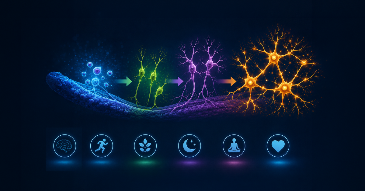
Why do some people stay mentally sharp well into their 80s and beyond, while others experience memory decline?
Scientists have been trying to answer this question for decades. A breakthrough new study published in Nature may offer an intriguing clue: the brains of older adults with exceptional memory, called superagers, continue to generate significantly more new neurons than their peers, and even much younger individuals. This discovery highlights a unique form of neurogenesis that may help explain why some brains stay resilient during aging. This discovery may actually be one of the superager brain secrets scientists have been hunting for.
Although researchers are still piecing together the full story, the findings could represent an important step toward understanding how the brain maintains cognitive health over time.
Medically reviewed by Kandace Licciardi, MD, Medical Director, Amen Clinics
A new, breakthrough study found that the brains of older adults with exceptional memory, called superagers, continue to generate significantly more new neurons than their peers, and even much younger individuals.
Superagers are individuals typically 80 years or older whose memory performance rivals people in their 50s and 60s. Structurally, their brains look younger as well with less shrinkage and fewer Alzheimer’s disease-type changes. Superagers are rare, accounting for less than 10 percent of the elderly population.
Northwestern University has been studying superagers for more than 25 years in their Northwestern University Superaging Research Program. Scientists have noted biological and behavioral differences in this group, including slower cortical thinning, greater gray matter volume, and stronger social engagement.
Superagers have better mental health, with less self-reported depression and anxiety, and fewer complaints of poor sleep than their peers, research has found.
Related: Healthy Brain at Every Age
In the study, led by scientists at University of Illinois Chicago, researchers examined post-mortem brain tissue from several groups:
The scientists focused on the hippocampus, a brain region essential for learning and memory. Using advanced genetic and molecular analysis techniques, they studied hundreds of thousands of cells from the hippocampus to understand how brain cell development differs between these groups.
One of the most striking findings involved neurogenesis.
For years, scientists debated whether the human brain continues producing new neurons during adulthood. Evidence has gradually suggested that it does, particularly in the hippocampus. This study provides new support for that idea and suggests that superagers may have a unique pattern of neurogenesis.
Researchers identified neural stem cells, neuroblasts, and immature neurons in the hippocampus. (One expert likens neural stem cells to babies, neuroblasts to teenagers, and immature neurons to near adults.) These indicate the early stages of brain cell development that eventually become mature neurons. Compared with typical aging brains, superagers showed a distinct neurogenesis profile, which the scientists described as a possible “resilience signature” in the brain.
Overall, superagers showed substantially more new neurons in their hippocampi. They had more of these immature neurons than young adults, roughly twice as many as normal adults their age, and about two and a half times more than those with Alzheimer’s disease.
In simple terms, superager brains appeared better able to generate new neurons and support their survival.
The hippocampus plays a major role in forming and storing memories.
In animal studies, newly generated neurons help integrate new information into memory circuits and support learning. When neurogenesis declines, memory function often declines as well. The new research suggests that in humans, maintaining neurogenesis may help preserve memory and cognitive abilities with age.
By contrast, the study also found that individuals with Alzheimer’s disease had fewer immature neurons, as well as disrupted neurogenesis pathways. This difference hints that the brain’s ability to generate new neurons could be an important factor in cognitive resilience.
Another key discovery involved the molecular signals that regulate neurogenesis.
The researchers examined neurogenic cells and found differences in gene activity and chromatin accessibility, which are mechanisms that control which genes turn on or off. These changes were found in two types of neurogenic cells: astrocytes and CA1 neurons.
These molecular patterns appeared to distinguish:
These findings suggest that these immature neurons have unique genetic and epigenetic characteristics that may make them resilient to aging, which may protect memory as people age.
The study does not mean scientists have discovered the secret to staying mentally young forever.
But it does suggest that ongoing neuron growth may be one important piece of the puzzle.
Understanding how the brain maintains neurogenesis could eventually lead to new strategies aimed at:
Researchers note that identifying the biological mechanisms behind this resilience could help guide future therapies designed to preserve brain function in aging populations.
Related: The 5-Minute Morning Routine to Boost Your Brain
Healthy aging is influenced by many factors, including genetics, lifestyle, brain health, and environmental exposures. There are many actions you can take to reduce your risk of Alzheimer’s disease and other types of dementia. This can include simple things like diet, improving social connections, and using methods to boost recollection.
This study offers an encouraging insight: some brains maintain the ability to renew themselves even in later life. If scientists can better understand how superagers’ brains achieve this, it could help unlock new ways to protect memory and cognitive function for more people as they age.
The research adds to a growing body of evidence that the brain remains more dynamic and adaptable than once believed. And that is good news for anyone interested in keeping their brain healthy for the long run!
A Superager is typically someone aged 80 or older whose memory performance is comparable to people decades younger. Researchers study them to understand why some brains resist age-related cognitive decline.
Neurogenesis is the process by which the brain produces new neurons (brain cells). In adults, this primarily occurs in the hippocampus, a region involved in memory and learning.
Although not everyone may reach superager status, research indicates that brain-healthy habits, such as staying socially connected, eating well, exercising, and keeping your mind active, can protect memory and slow cognitive decline. At Amen Clinics, we practice precision medicine, using brain SPECT imaging as part of our comprehensive evaluation to understand what is really happening in your brain. Our whole-body approach helps our clinicians develop personalized treatment plans that include natural ways to boost brain health and protect against cognitive decline, medication only when necessary, and other innovative therapies to help you strengthen your brain at any age.
Not yet. The research shows a strong association between increased neurogenesis and exceptional memory in superagers, but more studies are needed to confirm cause and effect.
The hippocampus is central to memory formation and learning. Changes in this region are commonly seen in aging and in conditions like Alzheimer’s disease.
Memory loss, dementia, and other mental health conditions can’t wait. At Amen Clinics, we practice precision medicine—using brain SPECT imaging and comprehensive evaluations to understand what’s really happening in your brain, not just your symptoms.
Our whole-body approach to holistic psychiatry combines cutting-edge neuroscience with natural ways to treat mental health conditions, including targeted nutrition, supplements, lifestyle strategies, therapy, and medications (when necessary). Every treatment plan is personalized to address the root causes of your struggles and support the health of your brain, body, and mind.
Don’t settle for guesswork. You deserve answers—and a plan built specifically for you. Speak with a Brain Health Advisor today at 888-288-9834 or visit our contact page to get started.
Disouky, A., Sanborn, M.A., Sabitha, K.R. et al. Human hippocampal neurogenesis in adulthood, ageing and Alzheimer’s disease. Nature (2026). https://doi.org/10.1038/s41586-026-10169-4
Garo-Pascual M, Gaser C, Zhang L, Tohka J, Medina M, Strange BA. Brain structure and phenotypic profile of superagers compared with age-matched older adults: a longitudinal analysis from the Vallecas Project. Lancet Healthy Longev. 2023 Aug;4(8):e374-e385. doi: 10.1016/S2666-7568(23)00079-X. Epub 2023 Jul 13. PMID: 37454673; PMCID: PMC10397152.
Toda T, Parylak SL, Linker SB, Gage FH. The role of adult hippocampal neurogenesis in brain health and disease. Mol Psychiatry. 2019 Jan;24(1):67-87. doi: 10.1038/s41380-018-0036-2. Epub 2018 Apr 20. PMID: 29679070; PMCID: PMC6195869.
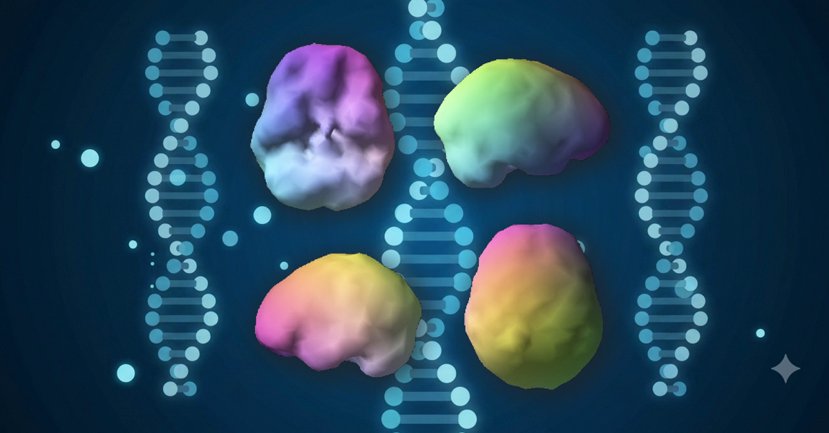
A massive new genetic study is challenging the way we define mental illness, and confirming that the brain is where the real story begins. The groundbreaking study, published in Nature, analyzed data from more than six million people and found that many psychiatric disorders share deep biological roots.
Researchers examined a total of fourteen conditions, including depression, anxiety, ADHD, bipolar disorder, schizophrenia, PTSD, autism, OCD, and substance use disorders. Instead of operating in isolated categories, these disorders appear to cluster together based on shared genetic patterns, particularly those related to brain development and synaptic function.
Significantly, the research suggests that mental illnesses are not simply a collection of siloed psychological symptoms, but overlapping biological systems that start in the brain, often as early as in utero.
For decades, psychiatric diagnoses have been based on symptom checklists rather than biology. This study reinforces what the brain-imaging work at Amen Clinics has shown for years: the current system of diagnosing mental health conditions is incomplete because it does not look directly at the organ it treats—the brain.
Medically reviewed by Rishi Sood, MD, Amen Clinics
New groundbreaking, genetic research suggests that mental illnesses are not simply a collection of siloed psychological symptoms, but overlapping biological systems that start in the brain, often as early as in utero.
The Nature study, published in December 2025, analyzed genetic data from more than six million people, including over one million individuals diagnosed with at least one psychiatric disorder. Researchers examined fourteen conditions, ranging from depression, anxiety, and PTSD to ADHD, autism, bipolar disorder, schizophrenia, OCD, and substance use disorders.
Instead of finding completely separate genetic pathways for each diagnosis, the team discovered that many of these disorders share common genetic risk factors. These shared risks clustered into broader biological groupings tied to brain development and synaptic function (how brain cells communicate with one another).
In other words, the same genetic variants can increase vulnerability to multiple mental health conditions. The study also found that certain traits, such as suicidality and loneliness, were genetically linked across several disorder clusters.
The findings suggest that psychiatric conditions may be better understood as overlapping brain-based systems rather than isolated categories, which counters the way mental illness has traditionally been classified.
Related: 3 Ways Brain SPECT Imaging Can Help Understand & Treat ADHD
The DSM-5, psychiatry’s diagnostic manual, groups conditions by clusters of symptoms. But symptoms don’t always tell the full story.
This new research identified genetic clusters that do not neatly align with traditional diagnostic labels. That means conditions like depression, bipolar disorder, and schizophrenia may share overlapping biological systems that begin affecting the brain very early in life.
In clinical practice, this mismatch happens frequently, according to Dr. Daniel Amen, founder of Amen Clinics, who was recently interviewed by Fox News regarding the new genetic study.
“We’ve had patients come in with the same diagnosis—say, depression—but when we scan their brains, we see wildly different patterns. One may have overactivity in the limbic system, another underactivity in the prefrontal cortex, and another damage from trauma or toxic exposure. They all feel depressed, but their brains tell different stories, and they each need a different approach to heal,” Dr. Amen told Fox News.
Dr. Amen explained further that diagnosing depression without looking at the brain is like diagnosing chest pain without examining the heart. Depression is a symptom, not a root cause.
This genetic mapping is an important step toward what many call “precision psychiatry.” It helps identify shared vulnerabilities and may eventually lead to earlier screening and prevention strategies.
However, genetics alone cannot determine outcomes. Association is not causation. Just because a gene is linked to a disorder does not mean it causes it, or that altering it will change the result.
Environment still matters. Your daily habits play a role in whether those genes are turned on or turned off. Genetics may load the gun, but stress, trauma, poor diet, infections, toxins, substance use, and head injuries often pull the trigger. On the other hand, a healthy lifestyle geared to support brain health can help reduce the risk of many mental health conditions.
That’s why a comprehensive brain health approach must look beyond DNA. SPECT imaging, which Amen Clinics uses in a comprehensive evaluation, bridges the gap between genetic risk and what is actually occurring in the brain.
“Genetics tells us what might happen. Imaging tells us what is happening right now,” Dr. Amen explained. Understanding what is happening in the brain leads to more accurate diagnoses, customized treatment plans and prevention programs, and better outcomes.
In the near future, genetic research may help refine subtypes of psychiatric disorders and improve early identification of risk. But it is not yet ready to guide day-to-day treatment decisions on its own.
Personalized psychiatry, however, is already possible. Brain SPECT imaging evaluates blood flow and activity patterns in the brain. Instead of guessing based on symptoms, imaging provides objective biological data that can be taken into consideration along with neuropsychological testing and clinical evaluations. Treatment can then be tailored to the individual brain pattern, not just the label written on a chart.
This approach moves mental health care from trial-and-error prescribing toward targeted, brain-based strategies that may include:
When care is personalized, outcomes improve.
If genetic research continues to advance alongside brain imaging, digital tracking tools, and neuroscience, the next couple of decades could dramatically reshape psychiatry.
“We’ll no longer be diagnosing based on symptoms alone. We’ll be diagnosing based on objective, biological data. Instead of calling someone ‘bipolar,’ we might say they have prefrontal cortex underactivity and temporal lobe instability with a genetic vulnerability in synaptic development, and then treat that specific pattern,” Dr. Amen commented further.
It’s possible that mental health care could shift from being reactive to proactive and preventive. “Imagine screening a 7-year-old for risk, optimizing their brain health early, and preventing future suffering,” Dr. Amen added.
And perhaps most importantly, stigma would decrease. That’s what happens naturally when mental health struggles are clearly understood as brain-based conditions, not character flaws. “If it’s your brain—not your fault—then there’s no shame, only opportunity for healing,” Dr. Amen explained.
This large-scale genetic study confirms something critical: mental health conditions share biological foundations rooted in brain function.
The future of care lies in combining genetic insights with real-time brain imaging and personalized treatment plans. That’s what Amen Clinics is already doing.
The large-scale study published in Nature found that many psychiatric disorders share overlapping genetic risk factors, particularly those related to brain development and synaptic function. Rather than operating as completely separate conditions, disorders such as depression, ADHD, bipolar disorder, schizophrenia, and anxiety appear to cluster together biologically. This challenges the traditional view that mental illnesses exist in isolated categories and supports a more brain-based understanding of psychiatric conditions.
Not necessarily, but it does suggest they may be incomplete. Current diagnoses are largely based on symptom checklists rather than biological data. Two people with the same diagnosis may have very different underlying brain patterns. The new research reinforces the need to move beyond symptom-based labels and toward evaluations that consider brain function, biology, and individual differences.
Genetics can increase vulnerability, but they do not determine destiny. Environmental factors such as stress, trauma, nutrition, sleep, toxins, substance use, and head injuries influence whether genetic risks are activated. A proactive, brain-healthy lifestyle can help reduce risk and improve resilience. Genetics may load the gun, but lifestyle and environment often determine the outcome.
The new genetic study found that many psychiatric disorders share overlapping biological roots tied to brain development and function. This supports what Amen Clinics’ brain imaging research has shown for decades: mental health conditions are brain-based, and symptom-only labels often miss the full picture.
Using SPECT imaging, Amen Clinics has observed that people with the same diagnosis can show very different brain activity patterns, while those with different diagnoses may share similar patterns. This overlap helps explain why conditions frequently co-occur and why treatment based solely on symptoms can fall short.
By looking directly at brain function, clinicians can better identify what is actually driving symptoms and create more personalized, targeted treatment plans.
Depression, anxiety disorders, and other mental health conditions can’t wait. At Amen Clinics, we provide personalized, science-backed treatment plans designed to target the root causes of your symptoms. Our 360-approach includes brain SPECT imaging, clinical evaluations, innovative therapeutic techniques, medications (when necessary), and holistic lifestyle recommendations to promote the health of your brain, body, and mind. Speak to a specialist today at 888-288-9834 or visit our contact page here.
Grotzinger, A.D., Werme, J., Peyrot, W.J. et al. Mapping the genetic landscape across 14 psychiatric disorders. Nature 649, 406–415 (2026). https://doi.org/10.1038/s41586-025-09820-3